. 2017 Aug;8(4):456.
doi: 10.4172/2157-7439.1000456. Epub 2017 Aug 31.

A Novel Nano-approach for Targeted Inner Ear Imaging

Affiliations

A Novel Nano-approach for Targeted Inner Ear Imaging

M N Kayyali et al. J Nanomed Nanotechnol. 2017 Aug.

Abstract

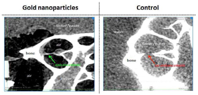
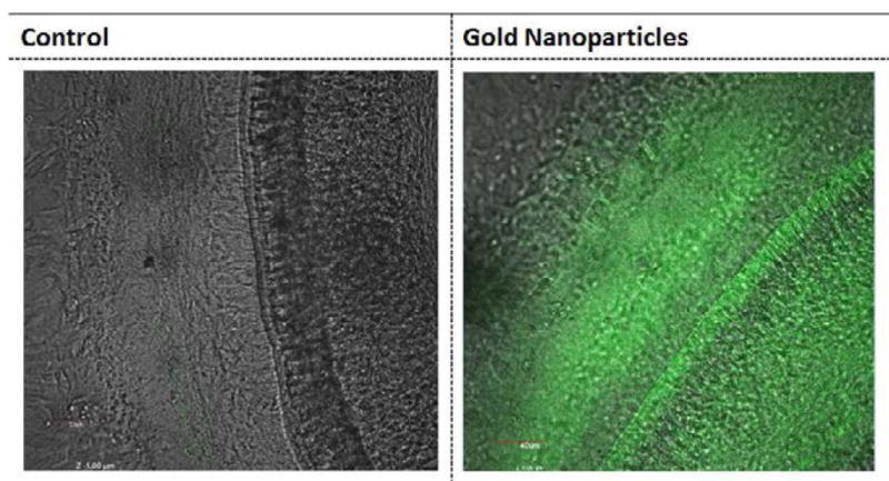
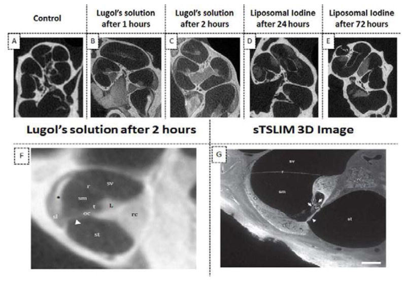
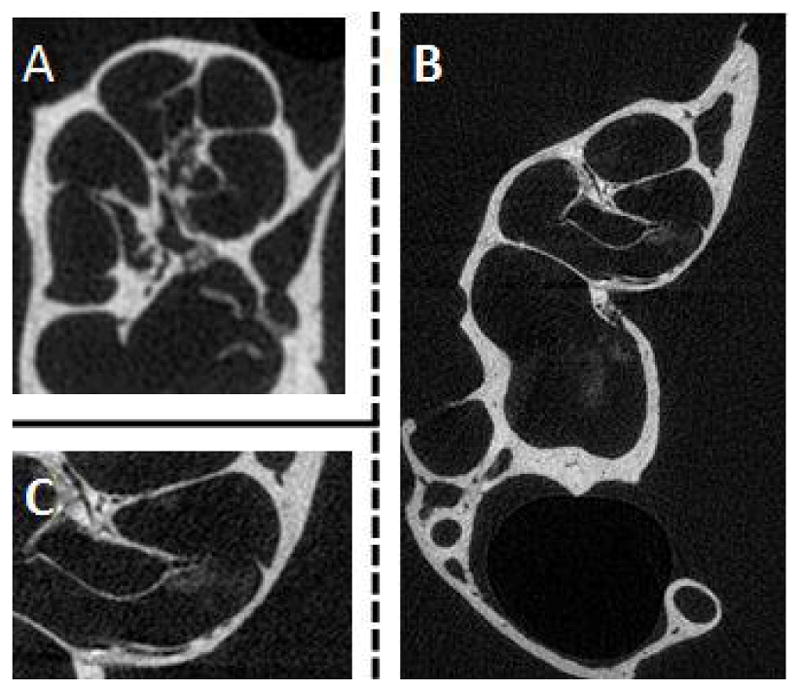
During the last decade, there have been major improvements in imaging modalities and the development of molecular imaging in general. However detailed inner ear imaging still provides very limited information to physicians. This is unsatisfactory as sensorineural hearing loss is the main cause of permanent hearing loss in adults and at least 134 genetic mutations that result in congenital hearing loss have been identified. We are still unable, in most cases where gross anatomical changes are not observed, to determine the exact cause of hearing loss at a cellular or molecular level in patients using non-invasive techniques. This limitation in inner ear diagnostic modalities is a major obstacle behind the delay in discovering treatments for many of the causes of sensorineural hearing loss. This paper initially investigated the use of targeted gold nanoparticles as contrast agents for inner ear imaging. These nanoparticles have many useful characteristics such as being easy to target and possessing minimal cytotoxicity. We were able to detect the nanoparticles diffusing in the hair cells using confocal microscopy. Regrettably, despite their many admirable characteristics, the gold nanoparticles were unable to significantly enhance CT imaging of the inner ear. Consequently, we investigated liposomal iodine as a potential solution for the unsatisfactory CT contrast obtained with the gold nanoparticles. Fortunately, significant enhancement of the micro-CT image was observed with either Lugol's solution or liposomal iodine, with Lugol's solution enabling fine inner ear structures to be detected.

Keywords: Gold nanoparticles; Inner ear imaging; Liposomal iodine; Targeted contrast agents.


Figures

Figure 1
Figure 1
Absorbance values for HEI-OC1 cells on a 6 day MTT time-course after exposure to GNPs. HEI-OC1 cells were incubated with three concentrations of GNPs: 0 (Green), 50 μM (red) and 100 μM (Blue) for up to 6 days. The Vybrant® MTT cell proliferation assay protocol was followed. This procedure was conducted in triplicate and demonstrated that GNPs have a minimal effect on cell viability.
Figure 2
Figure 2
Confocal microscopy images of mouse cochleae following treatment with GNPs (A) and the untreated control (B). GNPs were surgically applied into the mouse cochlea. After 24 hours, the cochleae were fixed and stained with a prestin-488 antibody. The results indicate that no significant difference in the OHC morphology was observed between cochleae that had been exposed to GNPs and the control cochleae. This strongly suggests that GNPs do not cause significant morphological changes.
Figure 3
Figure 3
Dark field microscopy images of HEI-OC1 cells. Targeted GNPs (0.25 mg/ml) (C) or untargeted GNPs (0.25 mg/ml) (B) were added to differentiated HEI-OC1 cells. The treated cells were then incubated for 24 hours, alongside control HEI-OC1 cells where GNPs were not added (A), to allow the nanoparticles to bind to the cells. The dark field images clearly show that much greater light scattering was observed with targeted GNPs (C) than with the other samples. This strongly suggests that the GNPs bound specifically to the HEI-OC1 cells: presumably to prestin.
Figure 4
Figure 4
Confocal microscopy images of cochlear organotypic cultures incubated with targeted GNPs (A), untargeted GNPs (B). Fluorescent GNPs (0.25 mg/ml) were added to cochlear organotypic cultures and the samples incubated for 24 hours to allow the nanoparticles to bind to the cells. The images clearly indicate that the targeted GNPs were binding specifically to the outer hair cells.
Figure 5
Figure 5
The use of gold nanoparticles to enhance CT imaging of the inner ear. GNPs were applied in vivo to mouse cochleae. The resulting CT images suggest that the presence of GNPs produced very limited enhancement of the CT image (green arrow), as compared to the tissue signal from the control sample where GNPs had not been added (red arrow).
Figure 6
Figure 6
Detection of gold nanoparticles inferred using confocal microscopy. GNPs bound to FITC were applied to mouse cochlea in vivo. After 24 hours, the cochleae were dissected and imaged under confocal microscopy. Fluorescence was observed throughout the cochlea that had been exposed to GNPs (B) while little fluorescence was detected in control cochlea where GNPs had not been added (A). This suggests that the GNPs injected into the mouse cochlear had fully diffused throughout the inner ear (B).
Figure 7
Figure 7
The use of iodine based contrast agents to enhance micro-CT images of mice cochleae. The ex vivo administration of Lugol’s iodine to cochleae produced significantly higher image signal and contrast at all time points than in images of untreated cochleae (A–C). In particular, there was a marked improvement in the imaging of the Organ of Corti (B and C). Most of the fine cochlear structures were observed in cochlear images taken after 2 hours incubation (F). The detail observable in the micro-CT image of (F) is comparable to that of a sTSLIM 3D optical image[40] (G): scala vestibuli (sv), media (sm) and tympani (st); basilar (arrowhead), tectorial (t) and Reissner’s membrane (r); stria vascularis (asterisk), spiral ligament (sl), organ of Corti (oc) with hair cells (arrows), spiral limbus (L) and Rosenthal’s canal (rc). The presence of liposomal iodine (LI) also enhanced the micro-CT images of the cochleae (D–E). Note: The micro-CT image in panel F was acquired at 12 micron resolution and is an average of 10 contiguous slices through a 120 micron thick slab.
Figure 8
Figure 8
Detection of liposomal iodine in cochlear CT imaging after in vivo application of liposomal iodine. Liposomal iodine was applied in vivo into mouse cochleae. After dissection, the micro-CT imaging system was used to scan the untreated control (A) and liposomal iodine-treated (B) cochleae. The magnified image of the liposomal iodine-treated cochlea (C) clearly shows the presence of the liposomal iodine, which had collected near the Organ of Corti. This was not detected in the control group (A). Although the liposomal iodine was detected inside the test cochlea, it did not enter the cells in sufficient quantities to enhance CT soft tissue imaging.

References

    1. Vio MM, Holme RH. Hearing loss and tinnitus: 250 million people and a US$10 billion potential market. Drug discovery today. 2005;10:1263–1265. - PubMed
    1. Shearer AE, DeLuca AP, Hildebrand MS, Taylor KR, Gurrola J, et al. Comprehensive genetic testing for hereditary hearing loss using massively parallel sequencing. Proceedings of the National Academy of Sciences of the United States of America. 2010;107:21104–92110. - PMC - PubMed
    1. Dror AA, Avraham KB. Hearing loss: mechanisms revealed by genetics and cell biology. Annu Rev Genet. 2009;43:411–437. - PubMed
    1. Joshi VM, Navlekar SK, Kishore GR, Reddy KJ, Kumar EC. CT and MR imaging of the inner ear and brain in children with congenital sensorineural hearing loss. Radiographics. 2012;32:683–698. - PubMed
    1. Han X, Ge R, Xie G, Li P, Zhao X, et al. Caspase-mediated apoptosis in the cochleae contributes to the early onset of hearing loss in A/J mice. ASN neuro 2015 - PMC - PubMed