. 2017 Mar;7(1):132-147.
doi: 10.1007/s12668-016-0303-0. Epub 2016 Oct 11.

Single-Molecule Interactions of a Monoclonal Anti-DNA Antibody with DNA

Affiliations

Single-Molecule Interactions of a Monoclonal Anti-DNA Antibody with DNA

Tatiana A Nevzorova et al. Bionanoscience. 2017 Mar.

Abstract

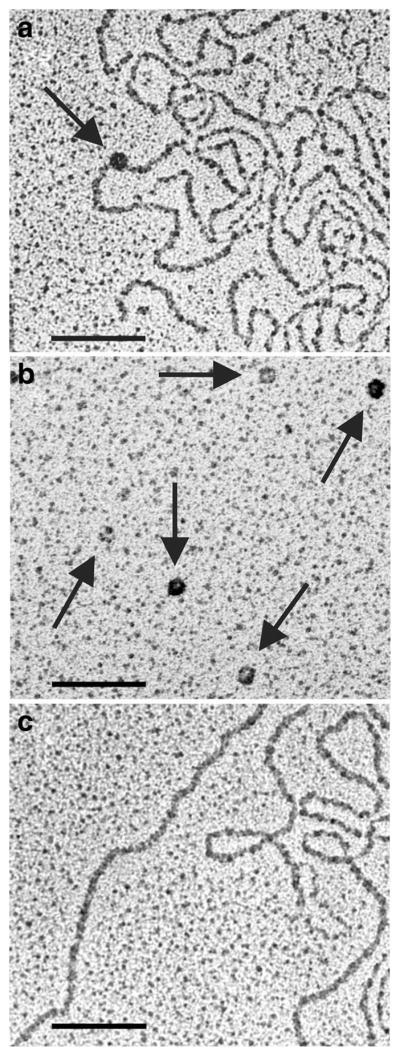
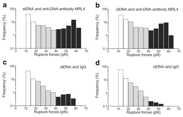
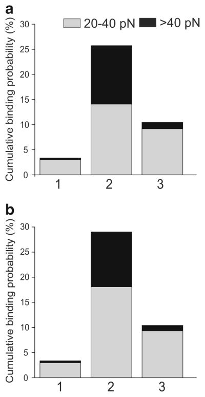
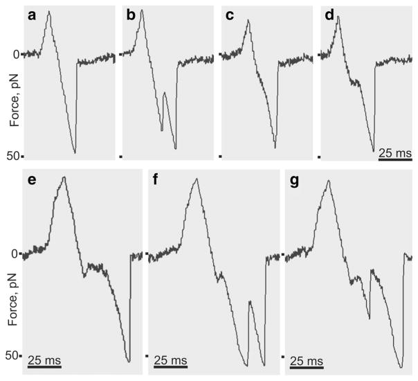
Interactions of DNA with proteins are essential for key biological processes and have both a fundamental and practical significance. In particular, DNA binding to anti-DNA antibodies is a pathogenic mechanism in autoimmune pathology, such as systemic lupus erythematosus. Here we measured at the single-molecule level binding and forced unbinding of surface-attached DNA and a monoclonal anti-DNA antibody MRL4 from a lupus erythematosus mouse. In optical trap-based force spectroscopy, a microscopic antibodycoated latex bead is trapped by a focused laser beam and repeatedly brought into contact with a DNA-coated surface. After careful discrimination of non-specific interactions, we showed that the DNA-antibody rupture force spectra had two regimes, reflecting formation of weaker (20-40 pN) and stronger (>40 pN) immune complexes that implies the existence of at least two bound states with different mechanical stability. The two-dimensional force-free off-rate for the DNA-antibody complexes was ~2.2 × 10⁻³ s-1, the transition state distance was ~0.94 nm, the apparent on-rate was ~5.26 s-1, and the stiffness of the DNA-antibody complex was characterized by a spring constant of 0.0021 pN/nm, suggesting that the DNA-antibody complex is a relatively stable, but soft and deformable macromolecular structure. The stretching elasticity of the DNA molecules was characteristic of single-stranded DNA, suggesting preferential binding of the MRL4 antibody to one strand of DNA. Collectively, the results provide fundamental characteristics of formation and forced dissociation of DNA-antibody complexes that help to understand principles of DNA-protein interactions and shed light on the molecular basis of autoimmune diseases accompanied by formation of anti-DNA antibodies.

Keywords: Anti-DNA antibody; DNA; Nanomechanics; Optical trap; Single-molecule force spectroscopy; Two-dimensional kinetics.


Figures

Fig. 1
Fig. 1
Electrophoresis of stDNA (a) and ctDNA (b) in a 0.6 % agarose gel stained with GelRed Nucleic Acid Gel Stain (Biotium, USA). The size of molecular weight markers (lower lanes) is shown in base pairs (bp). The plots over the gels represent optical density profiles for the DNA bands
Fig. 2
Fig. 2
PAAG (12.5 %) of purified monoclonal recombinant anti-DNA IgG MRL4. Stained by Coomassie R-250. Heavy chain (Hc) and light chain (Lc) of MRL4 are seen. Purity >95 %. The left line (MW) shows molecular weight markers
Fig. 3
Fig. 3
Schematic of the experimental model for investigation of single-molecule DNA-Ab interactions (not to scale). An Ab-coated latex bead was trapped by an oscillating focused laser beam and touched repeatedly to a stationary DNA-coated silica pedestal. A tensile force is generated when DNA and antibody bind while the bead moves away from the pedestal and get displaced from the focus of the beam
Fig. 4
Fig. 4
Immobilization of stDNA (a, b) and ctDNA (c, d) on clustered polyacrylamide-coated glutaraldehyde-activated silica pedestals. a and c show images of the DNA-coated pedestals in a bright field mode. b and d show fluorescent images of the same pedestals with covalently immobilized stDNA stained by Sibr Green II (b) and ctDNA with acridine orange (d). The fluorescence intensity of control images (DNA physisorption onto non-activated polyacrylamide silica pedestals and glutaraldehyde-activated polyacrylamide-coated pedestals without DNA) were negligible (not shown). Scale bar, 10 μm
Fig. 5
Fig. 5
a Typical AFM images of ctDNA–anti-DNA antibody MRL4 complex (arrows); b stDNA–MRL4 complex (arrows); c anti-DNA antibodies MRL4 alone (arrows, control). Scale bar, 100 nm
Fig. 6
Fig. 6
a Representative ctDNA–anti-DNA Ab complex (arrow); b isolated anti-DNA Abs (arrows); and c isolated ctDNA, visualized by transmission electron microscopy. Scale bar, 100 nm
Fig. 7
Fig. 7
Normalized rupture force histograms of non-specific interactions. a Control histogram of non-specifically interacting uncoated surfaces. Interactions of IgG-coated (b) and MRL4 anti-DNA Ab-coated (c) beads with uncoated pedestals. d Interactions of DNA-coated pedestals and uncoated beads. Signals that appeared as forces below 10 pN due to optical artifacts were considered nonbinding events or zero
Fig. 8
Fig. 8
Normalized rupture force histograms of the interactions of a monoclonal anti-DNA Ab MRL4 (a, b) and non-specific IgG (c, d) with stDNA (a, c) or ctDNA (b, d). Two force regimes are highlighted: the intermediate interactions at 20–40 pN (gray) and strong interactions >40 pN (black)
Fig. 9
Fig. 9
Average cumulative binding probabilities for various immunoglobulins interacting with stDNA (a) and ctDNA (b). The intermediate forces from 20 to 40 pN are in gray and strong forces >40 pN are in black. (1) Non-specific interactions (mean of all interacting surface pairs shown in Fig. 8); (2) a monoclonal anti-DNA Ab MRL4; (3) IgG, a control for non-immune interactions
Fig. 10
Fig. 10
Rupture force histograms (bars) and Bell’s model fits (curves) from two representative fitting exercises for the peaks of specific DNA-Ab forces >40 pN (inserts). a Fitting parameters are rF = 1200 pN/s, k0 = 0.0012 s−1, γ = 0.979 nm; b fitting parameters are rF = 1200 pN/s, k0 = 0.0026 s−1, γ = 0.91 nm
Fig. 11
Fig. 11
Data traces extracted from raw files showing various types of rupture force signals generated by the optical trap during forced unbinding of DNA-Ab complexes. The binding/unbinding events were detected as voltage signals calibrated in force units. Positive peaks reflect a compressive force exerted by a trapped bead on a pedestal upon touching. a Negative linear signal reflecting a one-step single-molecule unbinding event; b jagged two-step signal indicative of multiple interactions and stepwise unbinding; c a signal showing elastic elongation preceding unbinding; d signal with a rupture-force plateau (isometric stretching); e–g complex signals with a combination of unbinding scenarios: isometric stretching with elongation (e); isometric stretching and stepwise unbinding (f); isometric stretching, stepwise unbinding, and elastic elongation (g)
Fig. 12
Fig. 12
Fits of the force versus time data from the optical trap. The top two panels are for stDNA and the bottom two panels are for ctDNA. The curves were fitted to Eq. (5) using MATLAB’s curve fitting tool box. The values of Kuhn length obtained from the fits were close to the Kuhn lengths for single-stranded DNA

References

    1. Pisetsky DS. Antibody responses to DNA in normal immunity and aberrant immunity. Clinical and Diagnostic Laboratory Immunology. 1998;5:1–6. - PMC - PubMed
    1. Williams WM, Isenberg DA. A cross-sectional study of anti-DNA antibodies in the serum and IgG and IgM fraction of healthy individuals, patients with systemic lupus erythematosus and their relatives. Lupus. 1996;5:576–586. - PubMed
    1. Uchida K. Natural antibodies as a sensor of electronegative damage-associated molecular patterns (DAMPs) Free Radical Biology and Medicine. 2014;72:156–161. - PubMed
    1. Lora V, Bonaguri C, Gisondi P, Sandei F, Battistelli L, Russo A, et al. Autoantibody induction and adipokine levels in patients with psoriasis treated with infliximab. Immunologic Research. 2013;56:382–389. - PubMed
    1. Quan CP, Watanabe S, Pamonsinlapatham P, Bouvet J-P. Different dysregulations of the natural antibody repertoire in treated and untreated HIV-1 patients. Journal of Autoimmunity. 2001;17:81–87. - PubMed

LinkOut - more resources