

Effect of human amniotic epithelial cells on pro-fibrogenic resident hepatic cells in a rat model of liver fibrosis
- PMID: 29105277
- PMCID: PMC5783829
- DOI: 10.1111/jcmm.13396
Effect of human amniotic epithelial cells on pro-fibrogenic resident hepatic cells in a rat model of liver fibrosis
Abstract
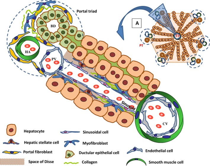
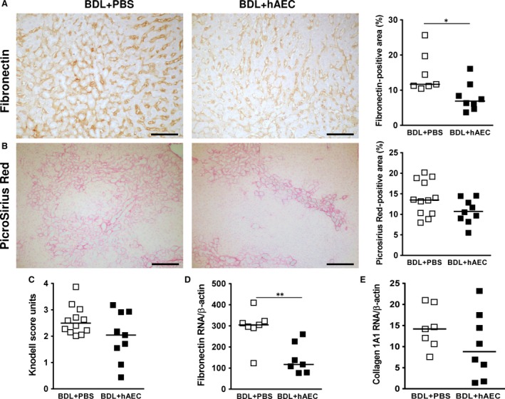
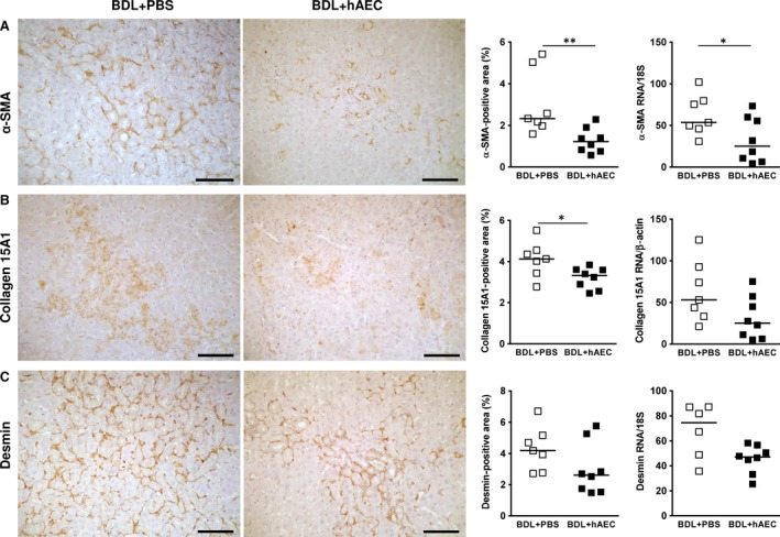
Myofibroblasts are key fibrogenic cells responsible for excessive extracellular matrix synthesis characterizing the fibrotic lesion. In liver fibrosis, myofibroblasts derive either from activation of hepatic stellate cells (HSC) and portal fibroblasts (PF), or from the activation of fibroblasts that originate from ductular epithelial cells undergoing epithelial-mesenchymal transition. Ductular cells can also indirectly promote myofibroblast generation by activating TGF-β, the main fibrogenic growth factor, through αvβ6 integrin. In addition, after liver injury, liver sinusoidal cells can lose their ability to maintain HSC quiescence, thus favouring HSC differentiation towards myofibroblasts. The amniotic membrane and epithelial cells (hAEC) derived thereof have been shown to decrease hepatic myofibroblast levels in rodents with liver fibrosis. In this study, in a rat model of liver fibrosis, we investigated the effects of hAEC on resident hepatic cells contributing to myofibroblast generation. Our data show that hAEC reduce myofibroblast numbers with a consequent reduction in fibronectin and collagen deposition. Interestingly, we show that hAEC strongly act on specific myofibroblast precursors. Specifically, hAEC reduce the activation of PF rather than HSC. In addition, hAEC target reactive ductular cells by inhibiting their proliferation and αvβ6 integrin expression, with a consequent decrease in TGF-β activation. Moreover, hAEC counteract the transition of ductular cells towards fibroblasts, while it does not affect injury-induced and fibrosis-promoting sinusoidal alterations. In conclusion, among the emerging therapeutic applications of hAEC in liver diseases, their specific action on PF and ductular cells strongly suggests their application in liver injuries involving the expansion and activation of the portal compartment.
Keywords: bile duct ligation; biliary liver fibrosis; ductular epithelial cells; human amniotic cells; human amniotic epithelial cells; human amniotic membrane; myofibroblasts; placenta-derived cells.
© 2017 The Authors. Journal of Cellular and Molecular Medicine published by John Wiley & Sons Ltd and Foundation for Cellular and Molecular Medicine.
Figures


Similar articles
-
Soluble factors derived from human amniotic epithelial cells suppress collagen production in human hepatic stellate cells.Cytotherapy. 2014 Aug;16(8):1132-44. doi: 10.1016/j.jcyt.2014.01.005. Epub 2014 Mar 15. Cytotherapy. 2014. PMID: 24642017
-
Transplantation of human amnion epithelial cells reduces hepatic fibrosis in immunocompetent CCl₄-treated mice.Cell Transplant. 2010;19(9):1157-68. doi: 10.3727/096368910X504496. Epub 2010 May 4. Cell Transplant. 2010. PMID: 20447339
-
The characteristics of activated portal fibroblasts/myofibroblasts in liver fibrosis.Differentiation. 2016 Sep;92(3):84-92. doi: 10.1016/j.diff.2016.07.001. Epub 2016 Aug 31. Differentiation. 2016. PMID: 27591095 Free PMC article. Review.
-
Role of TGF-β signaling in differentiation of mesothelial cells to vitamin A-poor hepatic stellate cells in liver fibrosis.Am J Physiol Gastrointest Liver Physiol. 2016 Feb 15;310(4):G262-72. doi: 10.1152/ajpgi.00257.2015. Epub 2015 Dec 23. Am J Physiol Gastrointest Liver Physiol. 2016. PMID: 26702136 Free PMC article.
-
Hepatic myofibroblasts: a heterogeneous population of multifunctional cells in liver fibrogenesis.Int J Biochem Cell Biol. 2009 Nov;41(11):2089-93. doi: 10.1016/j.biocel.2009.03.010. Epub 2009 Mar 31. Int J Biochem Cell Biol. 2009. PMID: 19782946 Review.
Cited by
-
Amniotic Membrane and Its Derivatives: Novel Therapeutic Modalities in Liver Disorders.Cells. 2023 Aug 21;12(16):2114. doi: 10.3390/cells12162114. Cells. 2023. PMID: 37626924 Free PMC article. Review.
-
Human Amniotic Epithelial Cells and Their Derived Exosomes Protect Against Cisplatin-Induced Acute Kidney Injury Without Compromising Its Antitumor Activity in Mice.Front Cell Dev Biol. 2022 Feb 3;9:752053. doi: 10.3389/fcell.2021.752053. eCollection 2021. Front Cell Dev Biol. 2022. PMID: 35186944 Free PMC article.
-
Mapping of the Human Amniotic Membrane: In Situ Detection of Microvesicles Secreted by Amniotic Epithelial Cells.Cell Transplant. 2023 Jan-Dec;32:9636897231166209. doi: 10.1177/09636897231166209. Cell Transplant. 2023. PMID: 37077027 Free PMC article.
-
Nanomaterials and hepatic disease: toxicokinetics, disease types, intrinsic mechanisms, liver susceptibility, and influencing factors.J Nanobiotechnology. 2021 Apr 16;19(1):108. doi: 10.1186/s12951-021-00843-2. J Nanobiotechnology. 2021. PMID: 33863340 Free PMC article. Review.
-
Arouse potential stemness: Intrinsic and acquired stem cell therapeutic strategies for advanced liver diseases.Cell Insight. 2023 Aug 11;2(5):100115. doi: 10.1016/j.cellin.2023.100115. eCollection 2023 Oct. Cell Insight. 2023. PMID: 37719773 Free PMC article. Review.
References
-
- Zatonski WA, Sulkowska U, Manczuk M, et al Liver cirrhosis mortality in Europe, with special attention to Central and Eastern Europe. Eur Addict Res. 2010; 16: 193–201. - PubMed
-
- Knittel T, Kobold D, Saile B, et al Rat liver myofibroblasts and hepatic stellate cells: different cell populations of the fibroblast lineage with fibrogenic potential. Gastroenterology. 1999; 117: 1205–21. - PubMed
-
- Lemoinne S, Cadoret A, Rautou PE, et al Portal myofibroblasts promote vascular remodeling underlying cirrhosis formation through the release of microparticles. Hepatology. 2015; 61: 1041–55. - PubMed
-
- Beaussier M, Wendum D, Schiffer E, et al Prominent contribution of portal mesenchymal cells to liver fibrosis in ischemic and obstructive cholestatic injuries. Lab Invest. 2007; 87: 292–303. - PubMed
Publication types
MeSH terms
Substances
LinkOut - more resources
Full Text Sources
Other Literature Sources
Medical
Research Materials
