Review
. 2018 Mar;7(2):10.1002/wdev.303.
doi: 10.1002/wdev.303. Epub 2017 Nov 3.

Limbal stem cells: identity, developmental origin, and therapeutic potential

Affiliations
Review

Limbal stem cells: identity, developmental origin, and therapeutic potential

Gabriel Gonzalez et al. Wiley Interdiscip Rev Dev Biol. 2018 Mar.

Abstract

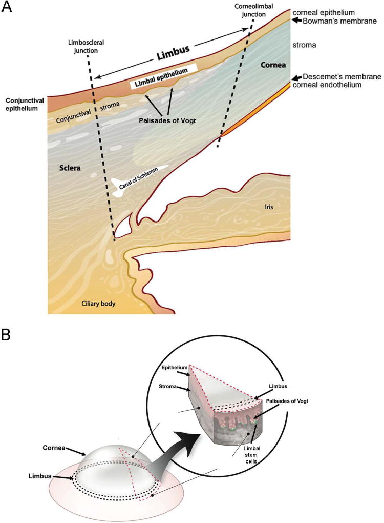
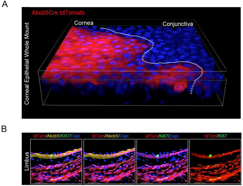
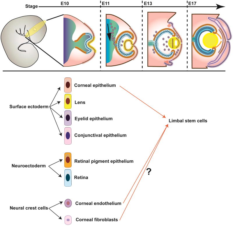
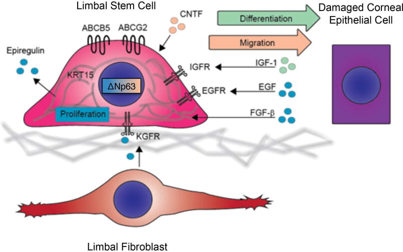
The cornea is our window to the world and our vision is critically dependent on corneal clarity and integrity. Its epithelium represents one of the most rapidly regenerating mammalian tissues, undergoing full-turnover over the course of approximately 1-2 weeks. This robust and efficient regenerative capacity is dependent on the function of stem cells residing in the limbus, a structure marking the border between the cornea and the conjunctiva. Limbal stem cells (LSC) represent a quiescent cell population with proliferative capacity residing in the basal epithelial layer of the limbus within a cellular niche. In addition to LSC, this niche consists of various cell populations such as limbal stromal fibroblasts, melanocytes and immune cells as well as a basement membrane, all of which are essential for LSC maintenance and LSC-driven regeneration. The LSC niche's components are of diverse developmental origin, a fact that had, until recently, prevented precise identification of molecularly defined LSC. The recent success in prospective LSC isolation based on ABCB5 expression and the capacity of this LSC population for long-term corneal restoration following transplantation in preclinical in vivo models of LSC deficiency underline the considerable potential of pure LSC formulations for clinical therapy. Additional studies, including genetic lineage tracing of the developmental origin of LSC will further improve our understanding of this critical cell population and its niche, with important implications for regenerative medicine. WIREs Dev Biol 2018, 7:e303. doi: 10.1002/wdev.303 This article is categorized under: Adult Stem Cells, Tissue Renewal, and Regeneration > Stem Cells and Disease Adult Stem Cells, Tissue Renewal, and Regeneration > Tissue Stem Cells and Niches Adult Stem Cells, Tissue Renewal, and Regeneration > Regeneration.


Figures

Figure 1
Figure 1. Schematic illustration of the human cornea and limbus
(A) Cross section illustrating the location and cellular layers of the cornea and limbus. (B) The Palisades of Vogt located at the corneo-limbal-scleral junction of the eye.
Figure 2
Figure 2. Contribution of ABCB5+ stem cells to corneal epithelial development and homeostasis
(A) Immunofluorescent image of corneal epithelium from a 1 month-old Abcb5/Cre/tdTomato mouse (whole-mount cornea). Abcb5/Cre transgenic mice were generated by insertion of an IRES-Cre cassette in the Abcb5 3’UTR downstream of the STOP codon located in exon 30. Abcb5/Cre mice were crossed with tdTomato (B6;129S6-Gt(ROSA)26Sortm9(CAG-tdTomato)Hze/J) mice for genetic lineage tracing studies. Whole-mount cornea preparations from Abcb5/Cre/tdTomato mice identified tdTomato-positive Abcb5-derived progeny cells within the entire adult mouse corneal epithelium. (B) Immunofluorescent (×60 magnification) images of tdTomato (red), Abcb5 (yellow) and Ki67 (green) co-expression in the limbus of Abcb5/Cre/tdTomato mice. The nuclei are stained with Dapi, 4′,6-diamidino-2-phenylindole (blue).
Figure 3
Figure 3. Developmental origin of LSC
(A) Schematic illustration of corneal development. From left to right: Extending neuroepithelium induces the surface ectoderm to form the lens, cornea, conjunctiva and eyelid. Migrating neural crest cells differentiate into corneal endothelium and stromal fibroblasts. (B) Contribution of diverse embryonic layers to the formation of the LSC niche. Current models suggest that LSC could be developmental descendants of the surface ectoderm as well as the periocular mesenchyme.
Figure 4
Figure 4. Current models of corneal epithelial migration and maintenance
(A) The Centripetal Migration Model, also known as the X-Y-Z hypothesis, of corneal regeneration. This model suggests that LSC located in the limbic region undergo centripetal migration and differentiation to maintain the corneal epithelium. (B) An alternative model suggests the existence of stem cells capable of corneal and conjunctival regeneration in the entire corneal epithelium. According to this model, the limbus represents a zone of equilibrium in which the expanding conjunctival and corneal epithelia are confronted in a mechanism reminiscent of tectonic plates; rupture of this equilibrium is suggested to result in migration of LSC onto the cornea.
Figure 5
Figure 5. Factors affecting corneal epithelial wound healing
Normal corneal wound healing is dependent on proliferation (blue), migration (orange) and differentiation (green) of corneal progenitors. Attenuated expression of LSC markers, including ABCB5, ABCG2, ΔNp63α or K15, is associated with abnormal corneal wound healing, which may result in increased corneal fragility, ulceration and clouding. Limbal epithelial cell proliferation is supported by expression of ΔNp63α and epiregulin in limbal basal epithelial cells, by keratinocyte growth factor (KGF) secreted from limbal fibroblasts, and by epidermal growth factor (EGF) and fibroblast growth factor-β (FGF-β) produced by damaged corneal epithelium. Migration is promoted by expression of ΔNp63α and ciliary neurotrophic factor (CNTF). Differentiation is induced by insulin-like growth factor-I (IGF-I), rapidly produced by injured corneal epithelium upon injury. KGFR, KGF receptor; EGFR, EGF receptor; IGFR, IGF receptor.

References

    1. Davanger M, Evensen A. Role of the pericorneal papillary structure in renewal of corneal epithelium. Nature. 1971;229(5286):560–1. - PubMed
    1. Schermer A, Galvin S, Sun TT. Differentiation-related expression of a major 64K corneal keratin in vivo and in culture suggests limbal location of corneal epithelial stem cells. J Cell Biol. 1986;103(1):49–62. - PMC - PubMed
    1. Tseng SC. Concept and application of limbal stem cells. Eye (Lond) 1989;3(Pt 2):141–57. - PubMed
    1. Lehrer MS, Sun TT, Lavker RM. Strategies of epithelial repair: modulation of stem cell and transit amplifying cell proliferation. J Cell Sci. 1998;111(Pt 19):2867–75. - PubMed
    1. Cotsarelis G, Cheng SZ, Dong G, Sun TT, Lavker RM. Existence of slow-cycling limbal epithelial basal cells that can be preferentially stimulated to proliferate: implications on epithelial stem cells. Cell. 1989;57(2):201–9. - PubMed

Publication types

MeSH terms

Substances