

Adipose Derived Stem Cells Affect miR-145 and p53 Expressions of Co-Cultured Hematopoietic Stem Cells
- PMID: 29105402
- PMCID: PMC5672106
- DOI: 10.22074/cellj.2018.4393
Adipose Derived Stem Cells Affect miR-145 and p53 Expressions of Co-Cultured Hematopoietic Stem Cells
Abstract
Objectives: Umbilical cord blood is used for transplantation purposes in regenerative medicine of hematological disorders. MicroRNAs are important regulators of gene expression that control both physiological and pathological processes such as cancer development and incidence. There is a new relation between p53 (tumor suppressor gene) and miR-145 (suppressor of cell growth) upregulation. In this study, we have assessed how adipose-derived stem cells (ADSCs) affect the expansion of hematopoietic stem cells (HSCs), as well as miR-145 and p53 expressions.
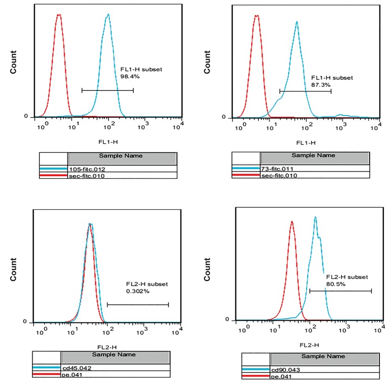
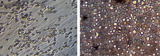
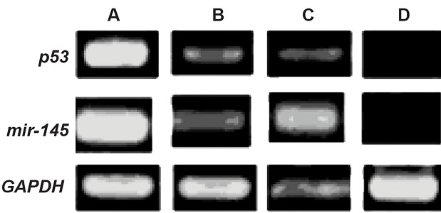
Materials and methods: In this experimental study, we cultured passage-3 isolated human ADSCs as a feeder layer. Flow cytometry analysis confirmed the presence of ADSC surface markers CD73, CD90, CD105. Ex vivo cultures of cordblood CD34+ cells were cultured under the following 4 culture conditions for 7 days: i. Medium only supplemented with cytokines, ii. Culture on an ADSCs feeder layer, iii. Indirect culture on an ADSCs feeder layer (Thin Cert™ plate with a 0.4 μm pore size), and iv. Control group analyzed immediately after extraction. Real-time polymerase chain reaction (PCR) was used to determine the expressions of the p53 and miR-145 genes. Flow cytometry analysis of cells stained by annexin V and propidium iodide (PI) was performed to detect the rate of apoptosis in the expanded cells.
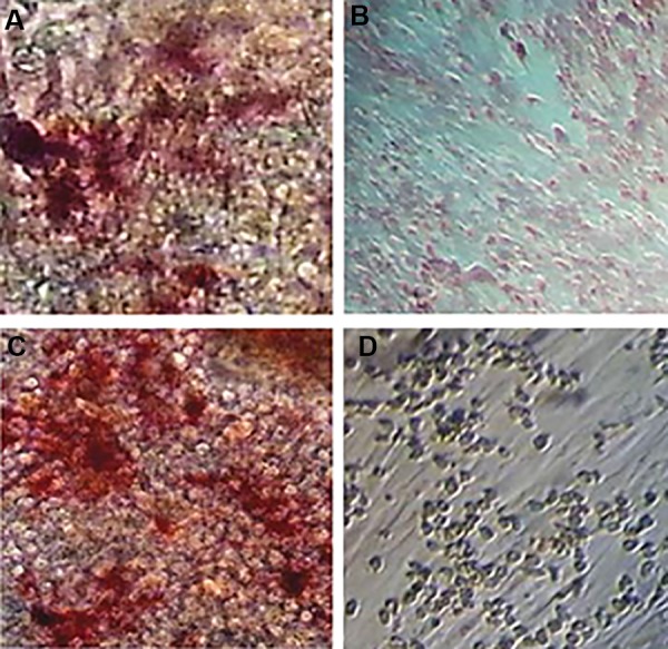
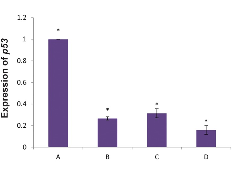
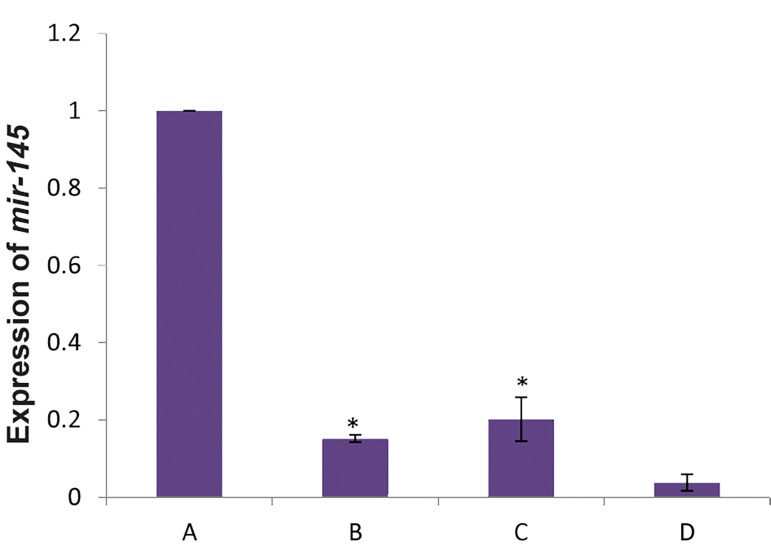
Results: ADSCs tested positive for mesenchymal stem cell (MSC) markers CD105, CD90, and CD73, and negative for HSC markers CD34 and CD45. Our data demonstrated the differentiation potential of ASCs to osteoblasts by alizarin red and alkaline phosphatase staining. MTT assay results showed a higher proliferation rate of CD34+cells directly cultured on the ADSCs feeder layer group compared to the other groups. Direct contact between HSCs and the feeder layer was prevented by a microporous membrane p53 expression increased in the HSCs group with indirect contact of the feeder layer compared to direct contact of the feeder layer. p53 significantly downregulated in HSCs cultured on ADSCs, whereas miR-145 significantly upregulated in HSCs cultured on ADSCs.
Conclusions: ADSCs might increase HSCs proliferation and self-renewal through miR-145, p53, and their relationship.
Keywords: Adipose Cell; Hematopoietic Stem Cell; MicroRNA.
Copyright© by Royan Institute. All rights reserved.
Conflict of interest statement
The authors declare no conflict of interest in this study.
Figures
References
-
- Walasek MA, van Os R, de Haan G. Hematopoietic stem cell expansion: challenges and opportunities. Ann N Y Acad Sci. 2012;1266:138–150. - PubMed
-
- Foroutan T. Increased miR33 expression in expanded hematopoietic stem cells cultured on adipose stem cells feeder layer. Iran J Ped Hematol Oncol. 2016;6(2):106–114.
-
- Saidi R, Rajeshkumar R, Shariftabrizi A, Zimmerman A, Walter O. Human adipose-derived mesenchymal stem cells promote liver regeneration. J Invest Surg. 2015;28(6):303–308. - PubMed
-
- Lindroos B, Suuronen R, Miettinen S. The potential of adipose stem cells in regenerative medicine. Stem Cell Rev. 2011;7(2):269–291. - PubMed
LinkOut - more resources
Full Text Sources
Research Materials
Miscellaneous