. 2017 Dec 1;127(12):4421-4436.
doi: 10.1172/JCI95545. Epub 2017 Nov 6.

Angiopoietin-1 is required for Schlemm's canal development in mice and humans

Affiliations

Angiopoietin-1 is required for Schlemm's canal development in mice and humans

Benjamin R Thomson et al. J Clin Invest. .

Abstract

Primary congenital glaucoma (PCG) is a leading cause of blindness in children worldwide and is caused by developmental defects in 2 aqueous humor outflow structures, Schlemm's canal (SC) and the trabecular meshwork. We previously identified loss-of-function mutations in the angiopoietin (ANGPT) receptor TEK in families with PCG and showed that ANGPT/TEK signaling is essential for SC development. Here, we describe roles for the major ANGPT ligands in the development of the aqueous outflow pathway. We determined that ANGPT1 is essential for SC development, and that Angpt1-knockout mice form a severely hypomorphic canal with elevated intraocular pressure. By contrast, ANGPT2 was dispensable, although mice deficient in both Angpt1 and Angpt2 completely lacked SC, indicating that ANGPT2 compensates for the loss of ANGPT1. In addition, we identified 3 human subjects with rare ANGPT1 variants within an international cohort of 284 PCG patients. Loss of function in 2 of the 3 patient alleles was observed by functional analysis of ANGPT1 variants in a combined in silico, in vitro, and in vivo approach, supporting a causative role for ANGPT1 in disease. By linking ANGPT1 with PCG, these results highlight the importance of ANGPT/TEK signaling in glaucoma pathogenesis and identify a candidate target for therapeutic development.

Keywords: Mouse models; Ophthalmology; Vascular Biology; endothelial cells.


Conflict of interest statement

Conflict of interest: S.E. Quaggin holds patents (US patent US9719135B2) related to therapeutic targeting of the ANGPT/TEK pathway in ocular hypertension and glaucoma and receives research support from, owns stock in, and is a director of Mannin Research.

Figures

Figure 1
Figure 1. ANGPT1 is the primary TEK ligand in SC development.
(A) Localization of Angpt1 expression in the limbal region. Cryosections from adult Angpt1GFP knockin mice show strong GFP expression in the TM and cells adjacent to the SC outer wall. Endomucin staining outlines the SC endothelium and capillaries of the superficial vascular plexus (SVP). (B) Angpt2 expression in the anterior chamber. Paraffin sections from adult X-gal–stained Angpt2LacZ knock-in mice show X-gal staining in the SC and corneal endothelium, as well as capillaries of the SVP. (C) In comparison with control littermates, eyes from mice with defective angiopoietin signaling failed to develop a normal SC. As previously described, Angpt1 Angpt2 double-knockout (Angpt1;2) mice completely lacked SC. Interestingly, however, while Angpt1WBΔE16.5 mice exhibited a severely hypoplastic canal with only focal development, Angpt2WBΔE16.5 mice had no apparent defect. (D) IOP measurements showed similar results (Angpt1 Angpt2 n = 12, control n = 42; Angpt1 n = 7, control n = 14; Angpt2 n = 3, control n = 4). This highlights the role of ANGPT1 as the major TEK ligand in canal development, though it suggests the possibility of limited compensation. Red arrows in C highlight SC. White arrows indicate the direction of the cornea. Note that faint staining in Angpt1;2 and Angpt1 knockout panels is out of focus light from the ciliary body and limbal vascular plexus. Scale bars: 50 μm (A and B) and 250 μm (C). ***P ≤ 0.001 as determined by Welch’s t test.
Figure 2
Figure 2. ANGPT1 is essential for SC formation.
SC development and maturation in Angpt1-knockout mice. Compared with littermate controls, SC development was disrupted in Angpt1WBΔE16.5 mice. Fewer CD31-positive sprouts emerge from the limbal capillaries, and these sprouts failed to consolidate and proliferate into a cohesive SC. In addition, PROX1 expression was dramatically reduced in the disorganized SC plexus of mutant mice, indicating that these cells failed to differentiate into the mature SC cell fate. Dashed lines in PROX1 panels highlight the CD31-positive sprout and SC area from matching CD31 staining. Littermate controls were used for all time points; eyes from 3 knockout and 3 control mice (P1, P12), 4 knockouts and 4 controls (P4), 6 knockouts and 5 controls (P7), 4 knockouts and 5 controls (P14), and 4 knockouts and 3 controls (P21) were analyzed. Three fields were captured per eye, and the results were averaged. AFU, background subtracted arbitrary fluorescence units. Scale bars: 50 μm; ×20 fields comprise an area of 65,536 μm2. *P ≤ 0.05, **P ≤ 0.01, ***P ≤ 0.001 as determined by Student’s t test.
Figure 3
Figure 3. SC and the TM are defective in Angpt1WBΔE16.5 mice.
(A) Iridocorneal angle region from control and Angpt1WBΔE16.5 mouse eyes obtained after imaging of plastic sections of eyes stained with H&E. Top panels show the location of SC and the TM within the iridocorneal angle. Magnified images of the region within the dotted box in the top panels are shown below for each panel. The control showed a well-developed SC and TM in contrast to mutants, in which the TM was hypoplastic and the SC was either absent or severely reduced in size. Control n = 4, Angpt1WBΔE16.5 n = 8. Scale bars: top panels, 50 μm, and bottom panels, 10 μm. (B) α-Smooth muscle actin–stained (α-SMA–stained) sections of the iridocorneal angle region from control and Angpt1WBΔE16.5 eyes, showing the ciliary muscle (CM) and TM. SC is indicated by a dashed oval. As in A, eyes lacking Angpt1 showed dramatic reduction in the size of SC and associated TM. Scale bar: 250 μm.
Figure 4
Figure 4. Angpt1-knockout mice exhibit clinical signs of glaucoma.
(A) Intraocular pressure (IOP) of Angpt1-knockout mice. Compared with control littermates, Angpt1WBΔE16.5 mice rapidly developed elevated IOP. Angpt1WBΔE16.5 n = 7, control n = 13. (B) Measurement of retinal ganglion cell number. When dissected at 19 weeks, mice lacking Angpt1 exhibited significant reductions in retinal ganglion cells compared with littermate controls. Angpt1WBΔE16.5 n = 5, control n = 4. **P ≤ 0.01, ***P ≤ 0.001 as determined by 2-way ANOVA followed by Bonferroni’s correction (A) or Student’s t test (B).
Figure 5
Figure 5. Pedigrees and ANGPT1 variants identified in 3 families.
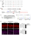
(A) Pedigrees of 3 families with ANGPT1 mutations. Specific mutations in ANGPT1 are listed below the different pedigrees, with carrier family members annotated as +/M. Affected individuals are indicated by solid black symbols. Note: White symbols do not exclude an undiagnosed late-onset disease. Parental genotypes/phenotypes are unknown for family 3. (B) Schematic representation of ANGPT1 protein domains and identified mutations in this study. S, signal peptide; SCD, superclustering domain; CCD, coiled-coil domain; L, linker domain; FLD, fibrinogen-like domain. (C) Sequence alignment of ANGPT1 K249 showing strong evolutionary conservation.
Figure 6
Figure 6. Expression and multimerization pattern of ANGPT1 variants.
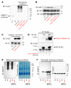
(A) Western blot of FLAG-tagged WT and variant ANGPT1 proteins secreted by transfected HEK293 cells. Under nonreducing conditions WT and ANGPT1K249R (K249R-FLAG) were observed in high-order oligomers. The premature stop codon upstream of the FLAG tag in ANGPT1Q236* (Q236*-FLAG) prevented production of full-length, FLAG-tagged protein. Surprisingly, however, despite the insertion of the FLAG tag upstream of the premature stop codon in ANGPT1R494* (R494_F498del-FLAG), no tagged protein was detected in the culture medium. (B) Western blot of HUVECs incubated with conditioned media from transfected HEK293 cells. Conditioned media from cells expressing WT ANGPT1 and ANGPT1K249R strongly induced AKT phosphorylation, while media from cells expressing ANGPT1Q236* and ANGPT1R494* did not. (C) Conditioned medium from HEK293 cells transfected with WT ANGPT1-FLAG or ANGPTK249R-FLAG was incubated with recombinant human TEK-Fc fusion protein to test the receptor-binding ability of this variant protein. (D) When coexpressed, FLAG-tagged WT ANGPT1 strongly pulled down ANGPT1Q236* with an HA tag inserted upstream of the premature stop codon (Q236_F498del-HA), indicating an interaction and potential dominant-negative activity. (E and F) Untagged WT and R494* variant ANGPT1 was expressed in HEK293 cells and immunoblotted using anti-ANGPT1 antibody. Only WT protein was detected in the conditioned medium, while ANGPT1R494* appears to be expressed but retained in the intracellular compartment. MG132 treatment was performed to block the proteasomal pathway. Coomassie blue staining and anti-tubulin (Tub) immunoblotting were used as loading controls for the conditioned media and cell lysate, respectively.
Figure 7
Figure 7. ANGPT1 p.R494* variant protein is trapped within intracellular aggregates.
(A and B) Subcellular localization of WT and R494* variant ANGPT1. When expressed in NIH 3T3 cells, WT ANGPT1-FLAG detected using anti-FLAG antibody showed a staining pattern consistent with localization in the ER (A) and Golgi apparatus (B). However, ANGPT1R494-FLAG was observed aggregated in intracellular vesicles positive for the ER marker PDI but not Golgin-97, a marker of the Golgi apparatus. (C) Interaction between WT and ANGPT1R494* within the cell. When coexpressed with untagged ANGPT1R494*, FLAG-tagged WT protein detected with anti-FLAG antibody was trapped in intracellular aggregates (white arrows) with untagged ANGPT1R494*. No WT ANGPT1-FLAG aggregates are observed in control cells coexpressing untagged WT ANGPT1. Scale bars: 10 μm.
Figure 8
Figure 8. Generation and analysis of Angpt1p.R494* mice.
(A) The CRISPR/Cas9 system was used to generate a novel ANGPT1R494*-expressing mouse line. While Arg-494 is conserved between mice and humans, it is encoded by different codons in each species. Therefore, different genomic DNA modifications are required for the p.R494* substitution in mice and humans. (B) Targeting strategy used to design guide RNAs. (C) Genotyping of N1 mice showing positive mutants following PCR and HpaII digestion. (D) Sanger sequencing analysis of a heterozygous founder mutant. (E) SC appears normal in Angpt1Null/WT and Angpt1p.R494*/WT heterozygous mice. In E, n = 3 WT/WT, 3 Null/WT, and 5 p.R494*/WT (SC area) or 3 (PROX1 expression) animals per group. Scale bars: 50 μm; ×20 fields comprise an area of 65,536 μm2.
Figure 9
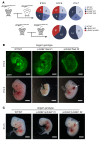
Figure 9. Angpt1p.R494* cannot replace WT Angpt1 in embryonic development.
(A) Heterozygous Angpt1p.R494*/WT mice were crossed with Angpt1Null/WT animals or inbred with Angpt1p.R494*/WT mice to generate p.R494*/Null or p.R494*/p.R494* offspring completely lacking WT ANGPT1 protein. While Angpt1p.R494*/Null pups were observed at normal Mendelian ratios early in embryonic development, double-mutant and homozygous mutant embryos died between E10.5 and E12.5 and no viable pups were born. (B and C) At E10.5, while all embryos were found alive, some Angpt1p.R494*/Null double mutants appeared smaller than control littermates and exhibited disorganized vasculature. By E12.5, all double-mutant and p.R494* homozygous embryos were found deceased (B and C), and some had been partially reabsorbed. Interestingly, some embryos had a visible hemorrhage in the region of the jugular lymph sac (white arrows), suggesting a possible defect in lymphovenous valve function. Pups found deceased. *No mortality was observed between birth and P14. Scale bars: 1 mm (B, top panels) and 2 mm (B, bottom panels, and C).
Figure 10
Figure 10. Angpt1p.R494* cannot replace WT Angpt1 in SC development.
Compared with littermate flox/flox and p.R494*/flox controls, at P21, Angpt1p.R494*/ΔE16.5 mice exhibited a hypomorphic SC with reduced PROX1 expression resembling that of Angpt1WBΔE16.5 mice. This finding confirms that the Angpt1p.R494* allele is functionally null in the context of SC development. n = 5 (flox/flox), 3 (p.R494*/flox and p.R494*/ΔE16.5), and 8 (WBΔE16.5) animals per group. **P < 0.01, ***P < 0.001 vs. flox/flox as determined by 1-way ANOVA followed by Bonferroni’s correction. AFU, background subtracted arbitrary fluorescence units. Scale bars: 50 μm; ×20 fields comprise an area of 65,536 μm2.
Figure 11
Figure 11. Defects in ANGPT/TEK signaling result in a hypoplastic SC insufficient for normal drainage.
(A) ANGPT1 is the primary TEK ligand in SC development, although limited compensation from ANGPT2 is possible. (B) Reductions in ANGPT1/TEK signaling lead to reduced angiogenic sprouting from the superficial vascular plexus (blue). The sparse sprouts observed in Angpt1WBΔE16.5 mice are unable to reach their neighbors, interdigitate, and mature into a normal SC (green), resulting in irregular canal formation, inadequate aqueous humor drainage, and glaucoma.

References

    1. Quigley HA, Broman AT. The number of people with glaucoma worldwide in 2010 and 2020. Br J Ophthalmol. 2006;90(3):262–267. doi: 10.1136/bjo.2005.081224. - DOI - PMC - PubMed
    1. Coleman AL, Miglior S. Risk factors for glaucoma onset and progression. Surv Ophthalmol. 2008;53(suppl 1):S3–S10. - PubMed
    1. Becker B. Tonography in the diagnosis of simple (open angle) glaucoma. Trans Am Acad Ophthalmol Otolaryngol. 1961;65:156–162. - PubMed
    1. Park DY, et al. Lymphatic regulator PROX1 determines Schlemm’s canal integrity and identity. J Clin Invest. 2014;124(9):3960–3974. doi: 10.1172/JCI75392. - DOI - PMC - PubMed
    1. Kizhatil K, Ryan M, Marchant JK, Henrich S, John SW. Schlemm’s canal is a unique vessel with a combination of blood vascular and lymphatic phenotypes that forms by a novel developmental process. PLoS Biol. 2014;12(7):e1001912. doi: 10.1371/journal.pbio.1001912. - DOI - PMC - PubMed

Publication types

MeSH terms