Review
. 2019 Jan:75-76:160-169.
doi: 10.1016/j.matbio.2017.10.007. Epub 2017 Oct 26.

Proteases and glycosidases on the surface of exosomes: Newly discovered mechanisms for extracellular remodeling

Affiliations
Review

Proteases and glycosidases on the surface of exosomes: Newly discovered mechanisms for extracellular remodeling

Ralph D Sanderson et al. Matrix Biol. 2019 Jan.

Abstract

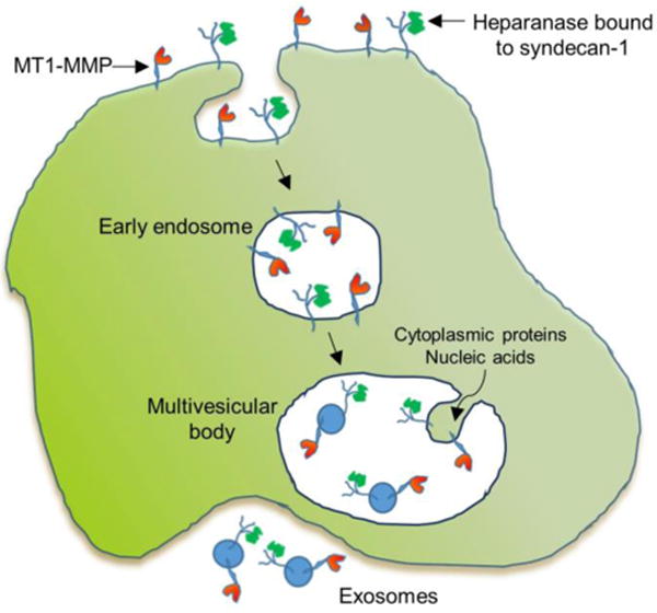
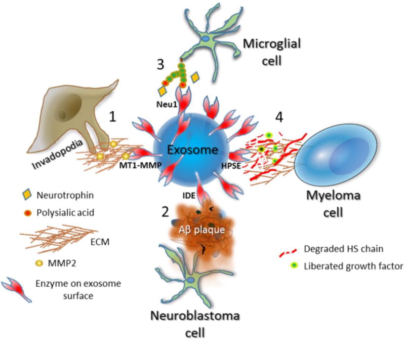
Emergence of the field of exosome biology has opened an exciting door to better understand communication between cells. These tiny nanovesicles act as potent regulators of biological function by delivering proteins, lipids and nucleic acids from the cell of origin to target cells. Recently, several enzymes including membrane-type 1 matrix metalloproteinase (MT1-MMP), insulin-degrading enzyme (IDE), sialidase and heparanase, among others, were localized on the surface of exosomes secreted by various cell types. These exosomal surface enzymes retain their activity and can degrade their natural substrates present within extracellular spaces. To date, enzymes on exosome surfaces have been associated with the mobilization of growth factors, degradation of extracellular matrix macromolecules and destruction of amyloid β plaques. This review focuses on the emerging role of exosomal surface enzymes and how this mechanism of remodeling within the extracellular space may regulate disease progression as related to cancer, inflammation and Alzheimer's disease.

Keywords: Alzheimer's; Cancer; Enzyme; Extracellular matrix; Extracellular vesicles.


Conflict of interest statement

Declarations of interest

The authors declare no competing financial interests.

Figures

Figure 1
Figure 1
This figure traces the route of MT1-MMP and heparanase through their endocytosis and subsequent transit through early endosome, multivesicular body and eventual secretion as surface components of the secreted exosomes. MT1-MMP localizes to the cell surface via its transmembrane domain; heparanase is retained on the cell surface through binding to the heparan sulfate chains of the transmembrane proteoglycan syndecan-1. Following membrane invagination and formation of the early endosome, the cell surface MT1-MMP and heparanase are present on the interior face of the early endosomal membrane. Within the multivesicular body, vesicles begin to form as the membrane invaginates and the forming vesicle fills with cytoplasmic contents (proteins, nucleic acids). Eventually the invaginations are pinched off to form vesicles followed by merging of the multivesicular body with the cell membrane and release of exosomes having MT1-MMP and heparanase on their surface.
Figure 2
Figure 2
Examples of enzymes on the exosome surface and their known functions. Depicted is an exosome having active enzymes on its surface and the functional impact of those enzymes within the extracellular space. The specific enzyme present depends on the cell that secreted the exosome. 1, MT1-MMP is on the surface of exosomes secreted at the site of invadopodia formation in breast and other cancers. MT1-MMP mediates shedding of cell surface molecules including syndecan-1 and CD44 and degrades fibrillar collagens, a host of other matrix molecules including fibronectin and vitronectin, and soluble molecules within the extracellular space. In addition, MT1-MMP activates MMP-2. Through this range of activities, MT1-MMP on the exosome surface may contribute to tumor migration and invasion as well as angiogenesis. 2, N2a neuroblastoma cells or BV-2 microglial cells secrete exosomes having a high level of insulin-degrading enzyme (IDE) on their surface. This enzyme is capable of degrading Aβ peptides that are prevalent in Alzheimer’s plaques. Statins enhance secretion of these exosomes containing high IDE levels and thus could prove therapeutically useful to attack plaques and diminish Alzheimer’s disease progression. 3, Microglial cells have high levels of polysialic acid on their surface. When stimulated by LPS, the cells rapidly enhance secretion of exosomes having the sialidase neuraminidase 1 (Neu1) on their surface. The sialidase cleaves the polysialic acid thereby liberating neurotrophic factors that are bound to the polysialic acid. The released neurotrophic factors have functional impact by regulating neural cell development and growth. 4, Myeloma cells expressing a high level of heparanase (HPSE) secrete exosomes having HPSE localized to the exosome surface. When added to ECM, the HPSE on the exosomes is capable of degrading heparan sulfate (HS) chains embedded within the ECM. This likely liberates growth factors bound to HS and by degrading ECM promotes tumor invasion and metastasis. Treatment of the myeloma cells with chemotherapeutic drugs enhances the secretion of exosomes loaded with HPSE as cargo leading to enhanced ECM degradation.

References

    1. Colombo M, Raposo G, Thery C. Biogenesis, secretion, and intercellular interactions of exosomes and other extracellular vesicles. Annu Rev Cell Dev Biol. 2014;30:255–89. - PubMed
    1. Kalluri R. The biology and function of exosomes in cancer. J Clin Invest. 2016;126:1208–15. - PMC - PubMed
    1. Yanez-Mo M, Siljander PR, Andreu Z, Zavec AB, Borras FE, Buzas EI, Buzas K, Casal E, Cappello F, Carvalho J, Colas E, Cordeiro-da Silva A, Fais S, Falcon-Perez JM, Ghobrial IM, Giebel B, Gimona M, Graner M, Gursel I, Gursel M, Heegaard NH, Hendrix A, Kierulf P, Kokubun K, Kosanovic M, Kralj-Iglic V, Kramer-Albers EM, Laitinen S, Lasser C, Lener T, Ligeti E, Line A, Lipps G, Llorente A, Lotvall J, Mancek-Keber M, Marcilla A, Mittelbrunn M, Nazarenko I, Nolte-’t Hoen EN, Nyman TA, O’Driscoll L, Olivan M, Oliveira C, Pallinger E, Del Portillo HA, Reventos J, Rigau M, Rohde E, Sammar M, Sanchez-Madrid F, Santarem N, Schallmoser K, Ostenfeld MS, Stoorvogel W, Stukelj R, Van der Grein SG, Vasconcelos MH, Wauben MH, De Wever O. Biological properties of extracellular vesicles and their physiological functions. J Extracell Vesicles. 2015;4:27066. - PMC - PubMed
    1. Raposo G, Stoorvogel W. Extracellular vesicles: exosomes, microvesicles, and friends. J Cell Biol. 2013;200:373–83. - PMC - PubMed
    1. Shimoda M, Khokha R. Proteolytic factors in exosomes. Proteomics. 2013;13:1624–36. - PubMed

Publication types

MeSH terms