Comparative Study
. 2018 Mar;14(3):352-366.
doi: 10.1016/j.jalz.2017.09.012. Epub 2017 Oct 31.

Conserved brain myelination networks are altered in Alzheimer's and other neurodegenerative diseases

Affiliations
Comparative Study

Conserved brain myelination networks are altered in Alzheimer's and other neurodegenerative diseases

Mariet Allen et al. Alzheimers Dement. 2018 Mar.

Abstract

Introduction: Comparative transcriptome analyses in Alzheimer's disease (AD) and other neurodegenerative proteinopathies can uncover both shared and distinct disease pathways.

Methods: We analyzed 940 brain transcriptomes including patients with AD, progressive supranuclear palsy (PSP; a primary tauopathy), and control subjects.

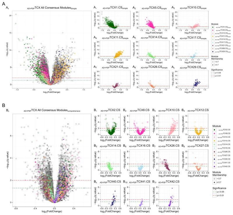
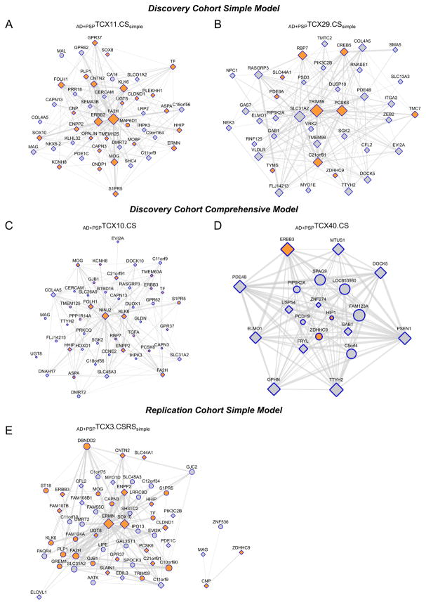
Results: We identified transcriptional coexpression networks implicated in myelination, which were lower in PSP temporal cortex (TCX) compared with AD. Some of these associations were retained even after adjustments for brain cell population changes. These TCX myelination network structures were preserved in cerebellum but they were not differentially expressed in cerebellum between AD and PSP. Myelination networks were downregulated in both AD and PSP, when compared with control TCX samples.

Discussion: Downregulation of myelination networks may underlie both PSP and AD pathophysiology, but may be more pronounced in PSP. These data also highlight conservation of transcriptional networks across brain regions and the influence of cell type changes on these networks.

Keywords: Alzheimer's disease; Cerebellum; Coexpression networks; Myelination; Progressive supranuclear palsy; Proteinopathies; Temporal cortex; Transcriptome.


Figures

Fig. 1
Fig. 1. Volcano plots of fold change vs. significance for differential gene expression (DGE) in the temporal cortex (TCX)
Results are shown for the primary analysis of AD vs. PSP TCX DGE in the Discovery Cohort, under the Simple (A0–A9) and Comprehensive (B0–B11) models. Each circle represents a transcript, which are colored differently according to the CEM they pertain to. Transcripts with strong module membership (MM) values≥0.7 are shown as filled circles; or empty circles if MM<0.7. Results are shown for all transcripts analyzed (A0, B0) and also separately for those CEM with consistent brain cell-enrichment across both models. DGE results that are significant at q≤0.05 or q≤0.01 are shown above the green and red dotted lines, respectively.
Fig. 2
Fig. 2. Oligodendrocyte networks in the Discovery and Replication Cohorts with disease association
Temporal cortex (TCX) oligodendrocyte-specific gene enriched networks in the Discovery Cohort under the Simple (A, B) and Comprehensive Models (C, D); and in the Replication Cohort under the Simple Model are shown for the primary AD vs. PSP analysis. These CEMs have significantly different levels between AD and PSP. None of the corresponding modules in the Replication Cohort under the Comprehensive Model were significantly associated with disease. The circles or squares represent the nodes for the genes within the CEM. For each module, the top 150 connections according to TOM weight are shown for genes with a MM > 0.7. The size of a node correlates with the number of connections for that node with others within the network. Gene transcripts that are enriched within oligodendrocytes are shown in orange. Transcripts with significant differential expression at q<0.05 are shown as a square. Thickness of the connection lines is determined by the weight of the connection.

References

    1. Goedert M. NEURODEGENERATION. Alzheimer’s and Parkinson’s diseases: The prion concept in relation to assembled Abeta, tau, and alpha-synuclein. Science. 2015;349:1255555. - PubMed
    1. Ertekin-Taner N, De Jager PL, Yu L, Bennett DA. Alternative Approaches in Gene Discovery and Characterization in Alzheimer’s Disease. Curr Genet Med Rep. 2013;1:39–51. - PMC - PubMed
    1. Cooper-Knock J, Kirby J, Ferraiuolo L, Heath PR, Rattray M, Shaw PJ. Gene expression profiling in human neurodegenerative disease. Nat Rev Neurol. 2012;8:518–30. - PubMed
    1. Tollervey JR, Wang Z, Hortobagyi T, Witten JT, Zarnack K, Kayikci M, et al. Analysis of alternative splicing associated with aging and neurodegeneration in the human brain. Genome Res. 2011;21:1572–82. - PMC - PubMed
    1. Miller JA, Oldham MC, Geschwind DH. A systems level analysis of transcriptional changes in Alzheimer’s disease and normal aging. J Neurosci. 2008;28:1410–20. - PMC - PubMed

Publication types