Comparative Study
. 2018 Jan 28:413:82-93.
doi: 10.1016/j.canlet.2017.10.029. Epub 2017 Oct 26.

Nimbolide reduces CD44 positive cell population and induces mitochondrial apoptosis in pancreatic cancer cells

Affiliations
Comparative Study

Nimbolide reduces CD44 positive cell population and induces mitochondrial apoptosis in pancreatic cancer cells

Sandeep Kumar et al. Cancer Lett. .

Abstract

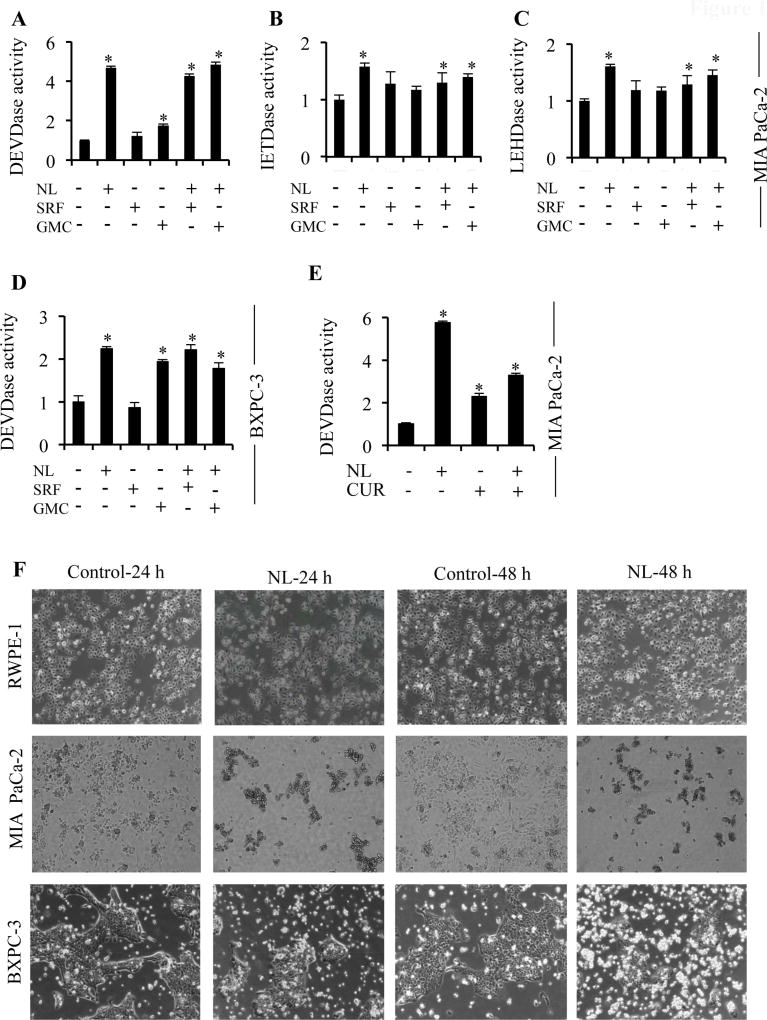
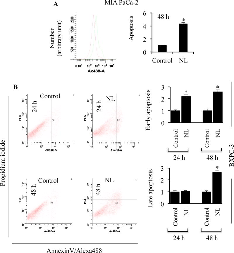
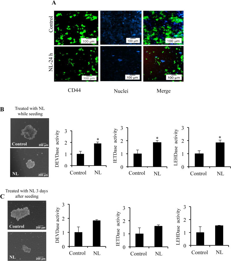
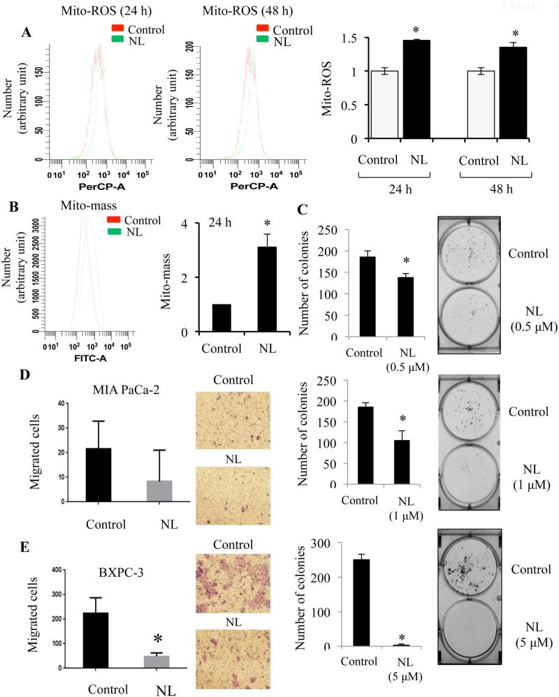
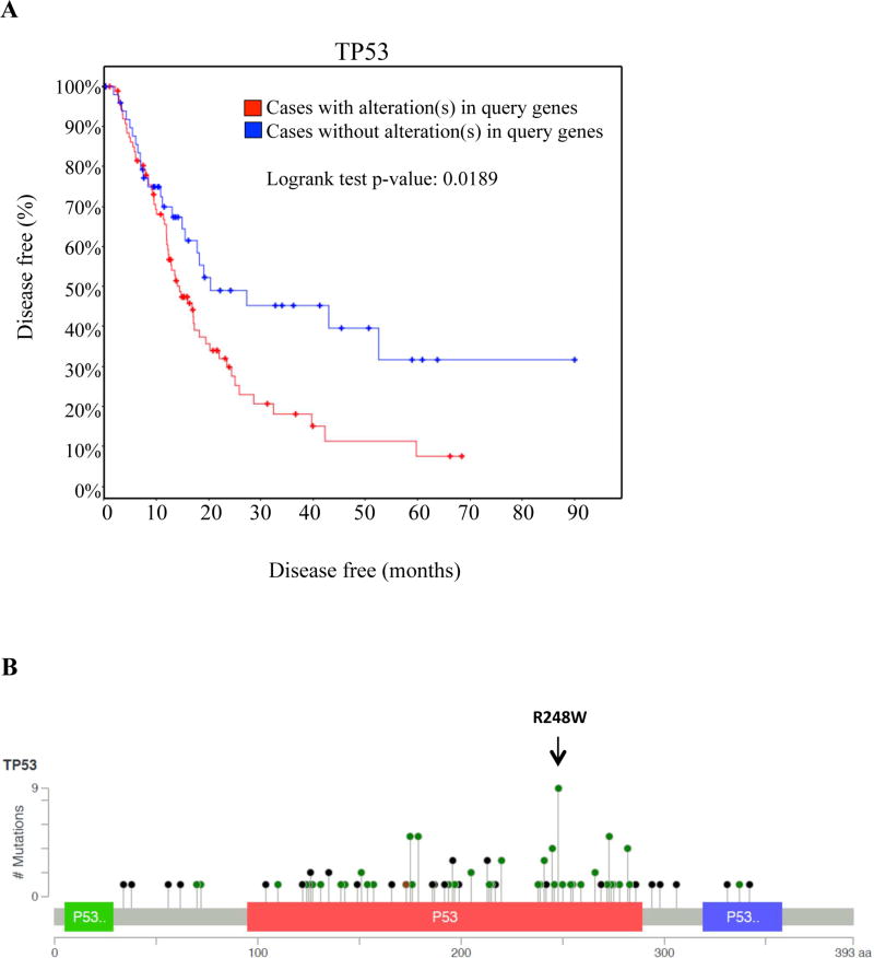
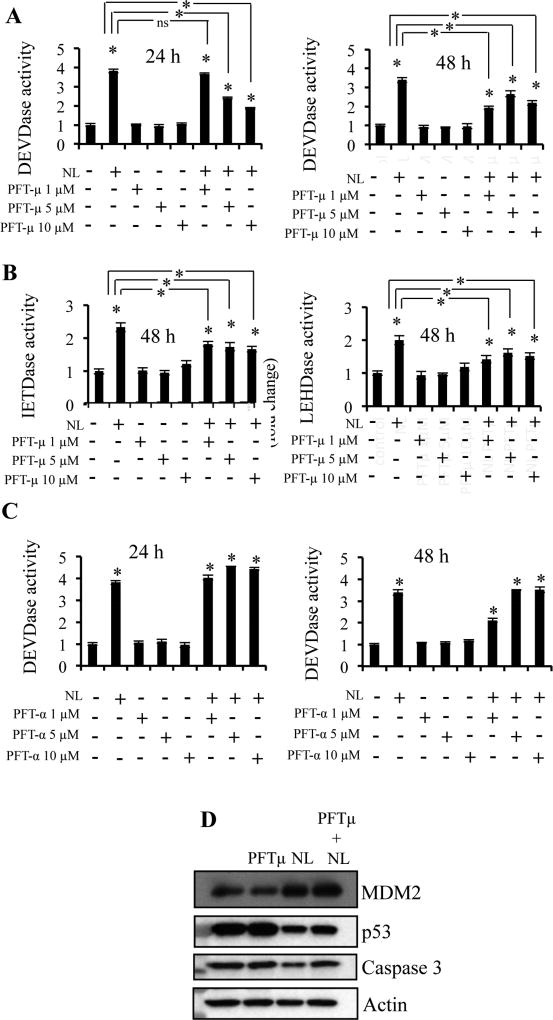
Pancreatic ductal adenocarcinoma (PDAC) is highly aggressive disease and current treatment regimens fail to effectively cure PDAC. Development of resistance to current therapy is one of the key reasons for this outcome. Nimbolide (NL), a triterpenoid obtained from Azadirachta indica, exhibits anticancer properties in various cancer including PDAC cells. However, the underlying mechanism of this anticancer agent in PDAC cells remains undefined. We show that NL exerts a higher level of apoptotic cell death compared to the first-line agent gemcitabine for PDAC, as well as other anticancer agents including sorafenib and curcumin. The anticancer efficacy of NL was further evidenced by a reduction in the CD44+ as well as cancer stem-like cell (CSC) population, as it causes decreased sphere formation. Mechanistically, the anticancer efficacy of NL associates with reduced mutant p53 as well as increased mitochondrial activity in the form of increased mitochondrial reactive oxygen species and mitochondrial mass. Together, this study highlights the therapeutic potential of NL in mutant p53 expressing pancreatic cancer.

Keywords: Apoptosis; Cancer stem cells; Mitochondria; Mutant p53; Nimbolide; Pancreatic cancer.


Conflict of interest statement

Conflict of interest: None

Figures

Figure 1
Figure 1. NL induces higher levels of caspase activity compared to current PDAC therapeutic agents
MIA PaCa-2 and BXPC-3 cells were treated with 5 µM NL, 5 µM Sorafenib (SRF), 5 µM Gemcitabine (GMC), or 5 µM Curcumin (CUR). (A–C) Caspase-3, -8 and -9 activities in MIA PaCa-2 cells after 24 h of NL, SRF, and GMC treatment. (D) Caspase-3 activity in BXPC-3 cells after 24 h of NL, SRF, and GMC treatment. (E) Caspase-3 activity in MIA PaCa-2 cells after 24 h of NL and CUR treatment. (F) Images of RWPE-1, MIA PaCa-2, and BXPC-3 cells treated with NL for 24 h and 48 h. Data are mean ± SD, n = 3 and presented as fold-change compared to respective controls. Statistical significance was determined by a t-test: *p<0.05.
Figure 2
Figure 2. NL induces apoptosis in pancreatic cancer cells
(A) Fold change in Annexin-V staining in MIA PaCa-2 cells following 5 µM NL exposure for 48 h. (B) Fold change in Annexin-V/PI staining in BXPC-3 cells following 5 µM NL exposure for 24 and 48 h. Data are mean ± SD, n = 3. Statistical significance was determined by a t-test: *p<0.05.
Figure 3
Figure 3. NL exposure reduces enrichment of cancer stem-like cells and inhibits sphere formation
(A) MIA PaCa-2 cells were treated with 5 µM NL and stained with FITC-tagged CD44 for 30 min. DAPI was used for nuclear staining. Expression of CD44+ populations at 24 h exposure of 5 µM NL was analyzed using fluorescence microscopy. For sphere formation, single cell suspensions of MIA PaCa-2 were seeded (1×10³ cells) into ultra-low attachment plates. Representative bright field images of spheres and caspase-3, -8 and -9 activities of spheres which were (B) treated on the day of seeding cells or (C) treated 3 days after seeding cells with 5 µM NL for 72 h. Data are mean ± SD, n = 3 and presented as fold-change compared to respective controls.. Statistical significance was determined by a t-test: *p<0.05.
Figure 4
Figure 4. NL enhances mitochondrial ROS and mass, inhibits colony formation, and impairs cell motility
BXPC-3 and MIA PaCa-2 cells were treated with NL (5 µM) for 24 and 48 h. Flow cytometry was carried out to estimate mitochondrial ROS (using MitoSOX red), and mitochondrial mass (using MitoTracker green). (A) Mitochondrial ROS (Mito-ROS) in BXPC-3 cells after 24 and 48 h of NL, and presented in fold-change. (B) Mitochondrial mass (Mito-mass) in MIA PaCa-2 cells after 24 h of NL, and presented in fold-change. (C) For clonogenic assays, single cell suspensions of MIA PaCa-2 cells were plated in six well plate (500 cells/well). After a 24 h incubation, cells were treated with 0.5 µM or 1 µM or 5 µM NL for 6 days. Following this, cells were fixed, stained with 0.5% crystal violet, and the number of colonies were quantified. For a transwell migration assay, MIA PaCa-2 and BXPC-3 cells were suspended in the upper chamber of a transwell insert (8 µm pore size) with media containing 0.5% FBS, while the lower chamber held media containing 7% FBS. Cells of the upper chamber received 5 µM NL and a 16 h incubation period was allowed. Non-migrated cells were removed. Cells which migrated through the insert were fixed with methanol and stained with 0.5% crystal violet. Using a light microscope at 20× magnification, the number of migrated cells per field were visualized and counted for (D) MIA PaCa-2 and (E) BX-PC3 cells. Data are mean ± SD, n = 3. Statistical significance was determined by a t-test: *p<0.05.
Figure 5
Figure 5. Mutations in p53 are prevalent in pancreatic adenocarcinoma and predict time to recurrence
(A) Time to recurrence between patients with no mutations (blue lines) and patients with p53 mutations (red line), displayed as a Kaplan-Meier graph. Censored values are presented as dots. (B) The distribution of frequently occurring p53 mutations along the amino acid sequence of p53. Note that the R248W alteration is the most prevalent in patients. Data was generated via cBioportal.
Figure 6
Figure 6. Reduction of mutant p53 associates with increased mitochondrial apoptosis
MIA PaCa-2 cells were plated. 24 h before NL exposure, cells were pretreated with 1, 5, or 10 µM Pifithrin-μ (PFT-μ) or Pifithrin-α (PFT-α) for p53 inhibition. Cells were then treated with 5 µM NL for 24 h. (A–B) Caspase-3, -8 and -9 activities in control and NL-treated MIA PaCa-2 cells previously treated with PFT-μ. (C) Caspase-3 activities in control and NL-treated MIA PaCa-2 cells previously treated with PFT-α. (D) Western Blot analysis of lysates from MIA PaCa-2 which were pretreated with 5 µM PFT-μ for 24 h, and then treated with 5 µM NL for 24 h. Data are mean ± SD, n = 3 and presented as fold-change compared to respective controls. Statistical significance was determined by a t-test: *p<0.05.

References

    1. Jemal A, Bray F, Center MM, Ferlay J, Ward E, Forman D. Global cancer statistics. CA Cancer J Clin. 2011;61(2):69–90. - PubMed
    1. Siegel RL, Miller KD, Jemal A. Cancer Statistics, 2017. CA Cancer J Clin. 2017;67(1):7–30. - PubMed
    1. Neoptolemos JP, Stocken DD, Friess H, Bassi C, Dunn JA, Hickey H, Beger H, Fernandez-Cruz L, Dervenis C, Lacaine F, Falconi M, Pederzoli P, Pap A, Spooner D, Kerr DJ, Buchler MW C. European Study Group for Pancreatic. A randomized trial of chemoradiotherapy and chemotherapy after resection of pancreatic cancer. N Engl J Med. 2004;350(12):1200–10. - PubMed
    1. Andersson R, Vagianos CE, Williamson RC. Preoperative staging and evaluation of resectability in pancreatic ductal adenocarcinoma. HPB (Oxford) 2004;6(1):5–12. - PMC - PubMed
    1. Canto MI, Harinck F, Hruban RH, Offerhaus GJ, Poley JW, Kamel I, Nio Y, Schulick RS, Bassi C, Kluijt I, Levy MJ, Chak A, Fockens P, Goggins M, Bruno M C. International Cancer of Pancreas Screening. International Cancer of the Pancreas Screening (CAPS) Consortium summit on the management of patients with increased risk for familial pancreatic cancer. Gut. 2013;62(3):339–47. - PMC - PubMed

Publication types

MeSH terms