. 2017 Nov;6(11):1339-1349.
doi: 10.1016/j.molmet.2017.08.010. Epub 2017 Sep 1.

The autonomic nervous system and cardiac GLP-1 receptors control heart rate in mice

Affiliations

The autonomic nervous system and cardiac GLP-1 receptors control heart rate in mice

Laurie L Baggio et al. Mol Metab. 2017 Nov.

Abstract

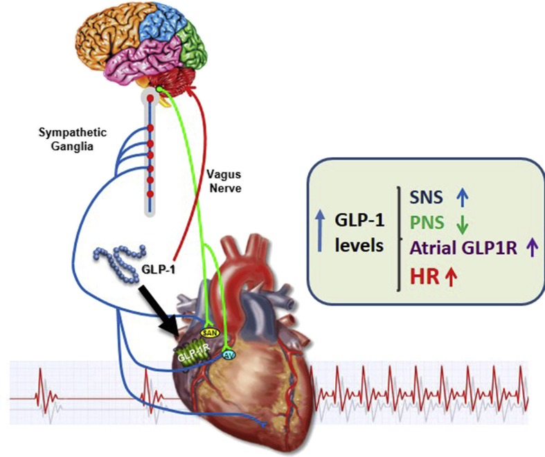
Objectives: Glucagon-like peptide-1 (GLP-1) is secreted from enteroendocrine cells and exerts a broad number of metabolic actions through activation of a single GLP-1 receptor (GLP-1R). The cardiovascular actions of GLP-1 have garnered increasing attention as GLP-1R agonists are used to treat human subjects with diabetes and obesity that may be at increased risk for development of heart disease. Here we studied mechanisms linking GLP-1R activation to control of heart rate (HR) in mice.

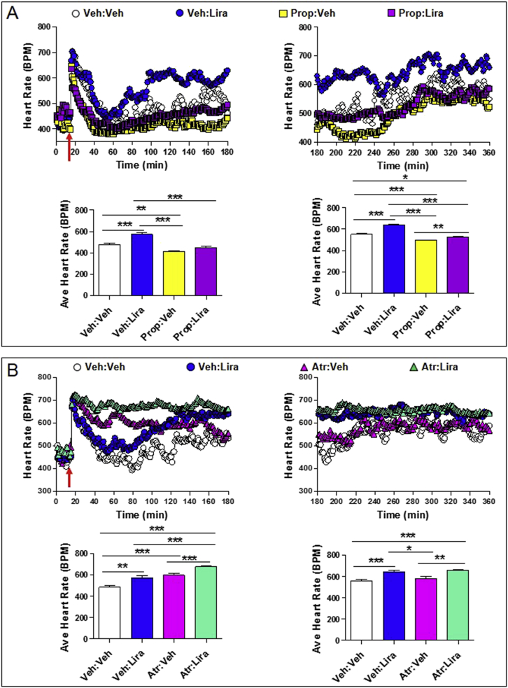
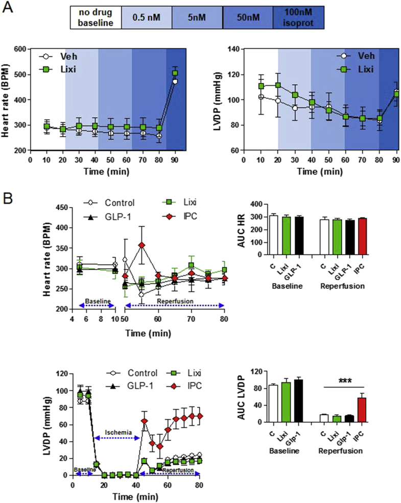
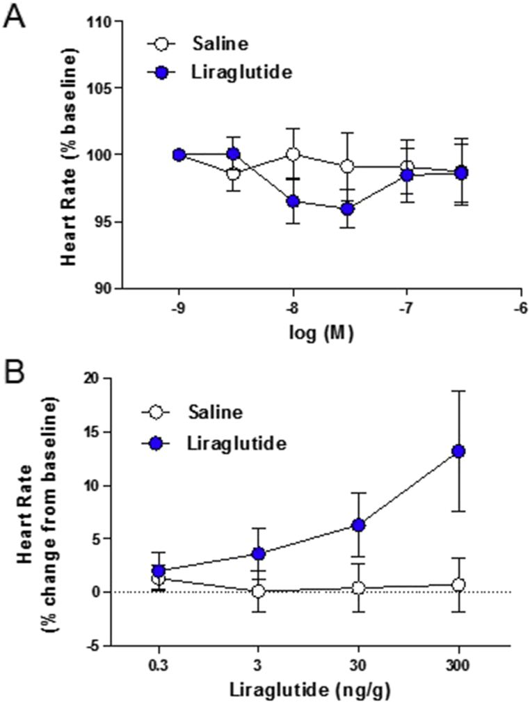
Methods: The actions of GLP-1R agonists were examined on the control of HR in wild type mice (WT) and in mice with cardiomyocyte-selective disruption of the GLP-1R (Glp1rCM-/-). Complimentary studies examined the effects of GLP-1R agonists in mice co-administered propranolol or atropine. The direct effects of GLP-1R agonism on HR and ventricular developed pressure were examined in isolated perfused mouse hearts ex vivo, and atrial depolarization was quantified in mouse hearts following direct application of liraglutide to perfused atrial preparations ex vivo.

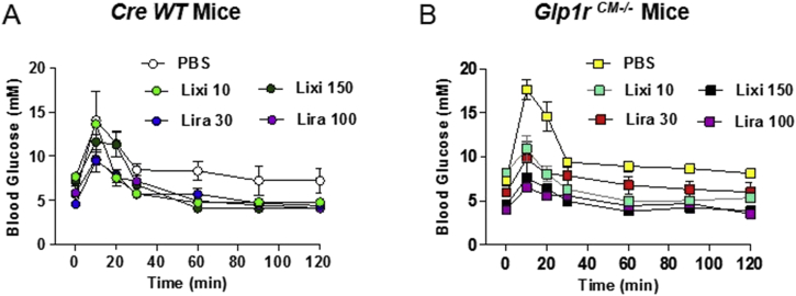
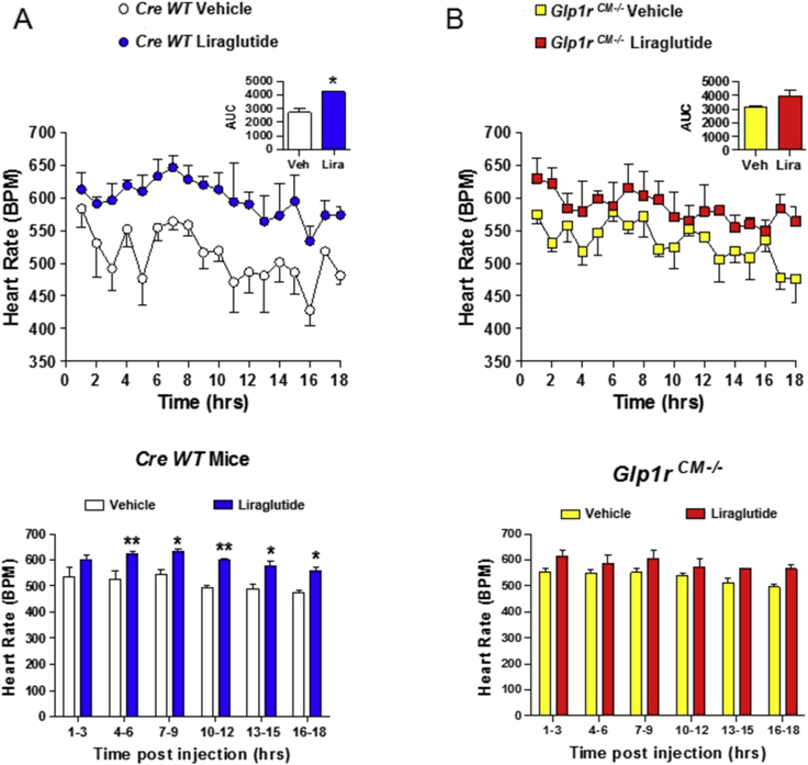
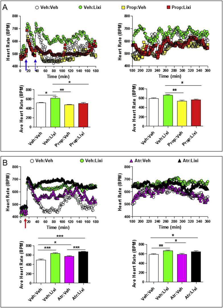
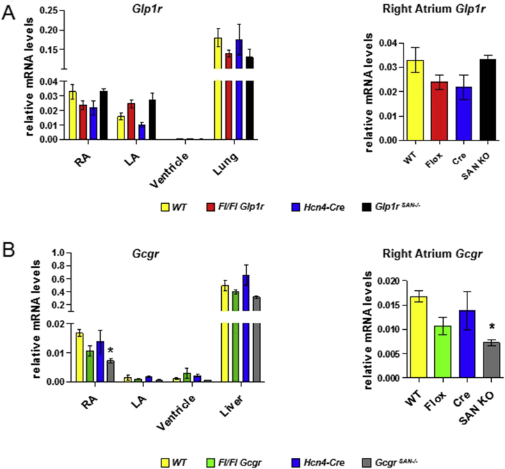
Results: Doses of liraglutide and lixisenatide that were equipotent for acute glucose control rapidly increased HR in WT and Glp1rCM-/- mice in vivo. The actions of liraglutide to increase HR were more sustained relative to lixisenatide, and diminished in Glp1rCM-/- mice. The acute chronotropic actions of GLP-1R agonists were attenuated by propranolol but not atropine. Neither native GLP-1 nor lixisenatide increased HR or developed pressure in perfused hearts ex vivo. Moreover, liraglutide had no direct effect on sinoatrial node firing rate in mouse atrial preparations ex vivo. Despite co-localization of HCN4 and GLP-1R in primate hearts, HCN4-directed Cre expression did not attenuate levels of Glp1r mRNA transcripts, but did reduce atrial Gcgr expression in the mouse heart.

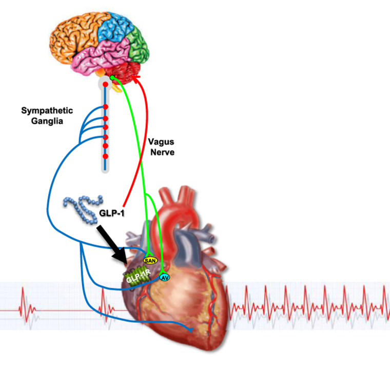
Conclusions: GLP-1R agonists increase HR through multiple mechanisms, including regulation of autonomic nervous system function, and activation of the atrial GLP-1R. Surprisingly, the isolated atrial GLP-1R does not transduce a direct chronotropic effect following exposure to GLP-1R agonists in the intact heart, or isolated atrium, ex vivo. Hence, cardiac GLP-1R circuits controlling HR require neural inputs and do not function in a heart-autonomous manner.

Keywords: Autonomic nervous system; Cardiac; Cardiovascular disease; Diabetes; GLP-1; Heart rate.


Figures

Image 1
Graphical abstract
Figure 1
Figure 1
Oral glucose tolerance in response to different doses of lixisenatide or liraglutide in cardiomyocyte-specific Glp1r knockout mice and controls. Blood glucose levels following oral glucose administration in Cre WT (A) and Glp1rCM−/− (B) mice treated with different doses of lixisenatide (Lixi), liraglutide (Lira), or vehicle (PBS). n = 2–4 mice/group.
Figure 2
Figure 2
Acute administration of liraglutide is associated with prolonged increases in heart rate in mice.Cre WT (A) and Glp1rCM−/− (B) mice were given a single ip injection of vehicle or liraglutide (30 μg/kg), and heart rate was recorded in conscious, freely moving mice for a total of 18 h (upper panels). All injections were administered 15 min after the start of data collection. The data in (A) and (B) are from 1 to 18 h post injection. The bar graphs (lower panels) represent the average heart rate at 3 h intervals following vehicle or liraglutide administration. Values are mean ± SE; n = 3 mice/group. *p < 0.05; **p < 0.01 vs. vehicle (Student's t-test of AUC data for upper panels; two-way ANOVA for lower panels). Body weights (g) are 39.9, 34.1, and 31.6 for Cre WT mice and 38.9, 31.7, and 32.3 for Glp1rCM−/− mice.
Figure 3
Figure 3
Autonomic nervous system-dependent effects of lixisenatide on heart rate. Heart rate (A and B; upper panels) and average heart rates (A and B; lower panels) over a 2–3 h interval in conscious, freely moving wild-type mice following a single ip injection of vehicle (Veh) or lixisenatide (Lixi; 10 μg/kg), in the presence or absence of propranolol (Prop; 5 mg/kg ip; sympathetic nervous system inhibitor) or atropine (Atr; 2 mg/kg ip; parasympathetic nervous system inhibitor). In (A), propranolol or vehicle (solid blue arrow; at time = 15 min) was administered 20 min prior to vehicle or lixisenatide (dotted blue arrow; at time = 35 min). In (B), atropine was administered at the same time as vehicle or lixisenatide (red arrow; at time = 15 min). For (A) and (B), data in panels on the left are from the first 3 h (0–180 min) of heart rate recordings and data in panels on the right are heart rate recordings from 3 to 6 h (180–360 min) after treatment administration. The average heart rate data for (A) and (B) lower left panels was calculated from 60 to 180 min or 40–180 min, respectively, when heart rates were stabilized following ip injections. Values are mean ± SE; n = 4–8 mice/group. Error bars have purposely been omitted from the heart rate data. *p < 0.05; **p < 0.01; ***p < 0.001 (One-way ANOVA). Body weights (g) are 31.0, 29.5, 30.8, and 30.0 for mice in (A) and 34.7, 36.3, 34.5, 34.7, 34.5, 32.4, 35.7, and 32.0 for mice in (B).
Figure 4
Figure 4
Autonomic nervous system-dependent effects of liraglutide on heart rate. Heart rate (A and B; upper panels) and average heart rate (A and B; lower panels) over a 2–3 h interval in conscious, freely moving wild-type mice following a single ip injection of vehicle (Veh) or liraglutide (Lira; 30 μg/kg), in the presence or absence of propranolol (Prop; 5 mg/kg ip; sympathetic nervous system inhibitor) or atropine (Atr; 2 mg/kg ip; parasympathetic nervous system inhibitor). In (A) and (B), propranolol or atropine was administered at the same time as vehicle or liraglutide (red arrow; at time = 15 min). For (A) and (B), data in panels on the left are from the first 3 h (0–180 min) of heart rate recordings and data in panels on the right are from 3 to 6 h (180–360 min) after treatment administration. The average heart rate data for (A) and (B) lower left panels was calculated from 60 to 180 min or 40–180 min, respectively, when heart rates were stabilized following ip injections. Values are mean ± SE; n = 5–8 mice/group. Error bars have been purposely omitted from the heart rate data. *p < 0.05; **p < 0.01; ***p < 0.001 (One-way ANOVA). Body weights (g) are 33.0, 32.0, 30.0, 34.0, and 30.0 for mice in (A) and 34.5, 36.6, 34.0, 35.2, 35.0, 32.2, 35.6, and 32.2 for mice in (B).
Figure 5
Figure 5
Glp1r mRNA levels are not reduced in floxed Glp1r mice expressing HCN4-Cre.Glp1r (A) and Gcgr (B) mRNA levels were measured in right atria (RA), left atria (LA), ventricle and lung or liver from wild-type control (WT), Glp1rflox/flox (Fl/Fl Glp1r), Gcgrflox/flox (Fl/Fl Gcgr), Hcn4-Cre and sinoatrial node (SAN)-specific Glp1r (Glp1rSAN KO) or Gcgr (GcgrSAN KO) knockout mice. Panels on the right are an expanded view of Glp1r or Gcgr mRNA expression in the right atrium which contains the sinoatrial node. Values are mean ± SE; n = 3–8 mice/group. *p < 0.05 vs. WT (One-way ANOVA).
Figure 6
Figure 6
Lixisenatide has no effect on heart rate or LVDP in isolated mouse hearts ex vivo. (A) Heart rate and left ventricular developed pressure (LVDP) in aerobic perfused ex vivo mouse hearts exposed to vehicle (Veh) or increasing doses of lixisenatide (Lixi). (B) Analysis of heart rate (upper panels) and LVDP (lower panels), at baseline and during reperfusion, following a 30 min period of no-flow global ischemia in ex vivo isolated mouse hearts that were exposed to vehicle (control), 0.5 nM GLP-1 7-36 (GLP-1), 5 nM lixisenatide (Lixi), or ischemia preconditioning (IPC). Heart rate recordings during the ischemic period (upper left panel) are very high due to quivering of the heart and have been omitted from the graph. The panels on the right depict AUC data for heart rate or LVDP at baseline and during the reperfusion period. In (A), isoproterenol (isoprot) was used as a positive control at the end of each perfusion. Values are mean ± SE; n = 5–11 hearts/group. ***p < 0.001 for IPC vs all other treatments (One-way ANOVA of AUC LVDP data).
Figure 7
Figure 7
Effects of liraglutide on depolarization frequency of isolated atrial preparations and HR control in anaesthetized mice. (A) Atrial preparations from wild type mice were isolated, loaded with a voltage-sensitive fluorescent dye (di-4-ANEPPS), superfused, and imaged at 1000 frames/sec to determine baseline atrial beating rates (heart rate) as described in the Methods. The indicated doses of liraglutide or saline vehicle were superfused to examine their effects on baseline atrial depolarization frequencies. Data are expressed as the mean % ± SE of the baseline heart rate values for each group (saline baseline; 414 ± 27 bpm, liraglutide baseline; 411 ± 28 bpm). n = 17 atrial preparations/group. At the end of these experiments, atria were subsequently exposed to isoproterenol, which increased sinoatrial node firing rates by more than 200 b.p.m. (B) Heart rate was measured in isoflurane-anaesthetized wild type mice administered the indicated dose of liraglutide or saline. Data are expressed as the mean percentage increase ± SE above basal heart rate prior to drug administration. n = 10 mice/group. For (A) and (B), experimenters were blinded to control and drug group identities until after data processing and analysis.
Figure 8
Figure 8
Direct and indirect mechanisms linking GLP-1R signaling to control of heart rate. GLP-1 engages GLP-1 receptors in the central, peripheral, and autonomic nervous systems to enhance sympathetic nervous system (SNS) activity, and reduce parasympathetic nervous system (PNS) activity. GLP-1 additionally activates atrial GLP-1 receptors which also contribute, with inputs from the SNS and PNS, to control of heart rate.

References

    1. Burmeister M.A., Ayala J.E., Smouse H., Landivar-Rocha A., Brown J.D., Drucker D.J. The hypothalamic glucagon-like peptide 1 receptor is sufficient but not necessary for the regulation of energy balance and glucose homeostasis in mice. Diabetes. 2017;66(2):372–384. - PMC - PubMed
    1. Drucker D.J. The cardiovascular biology of glucagon-like Peptide-1. Cell Metabolism. 2016;24(1):15–30. - PubMed
    1. Stamler J., Vaccaro O., Neaton J.D., Wentworth D. Diabetes, other risk factors, and 12-yr cardiovascular mortality for men screened in the Multiple Risk Factor Intervention Trial. Diabetes Care. 1993;16(2):434–444. - PubMed
    1. Zinman B., Wanner C., Lachin J.M., Fitchett D., Bluhmki E., Hantel S. Empagliflozin, cardiovascular outcomes, and mortality in type 2 diabetes. New England Journal of Medicine. 2015;373(22):2117–2128. - PubMed
    1. Barragan J.M., Rodriguez R.E., Blazquez E. Changes in arterial blood pressure and heart rate induced by glucagon-like peptide-1-(7-36 amide) in rats. American Journal of Physiology. 1994;266:E459–E466. - PubMed

Publication types

MeSH terms

LinkOut - more resources