. 2017 Nov 6:23:5283-5293.
doi: 10.12659/msm.907449.

Klotho Improves Cardiac Function by Suppressing Reactive Oxygen Species (ROS) Mediated Apoptosis by Modulating Mapks/Nrf2 Signaling in Doxorubicin-Induced Cardiotoxicity

Affiliations

Klotho Improves Cardiac Function by Suppressing Reactive Oxygen Species (ROS) Mediated Apoptosis by Modulating Mapks/Nrf2 Signaling in Doxorubicin-Induced Cardiotoxicity

Huolan Zhu et al. Med Sci Monit. .

Abstract

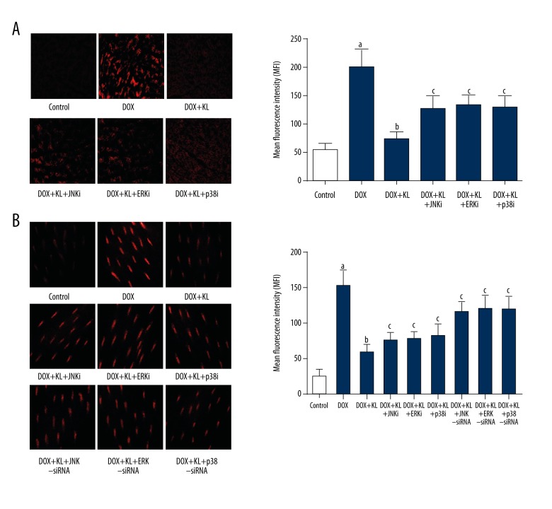
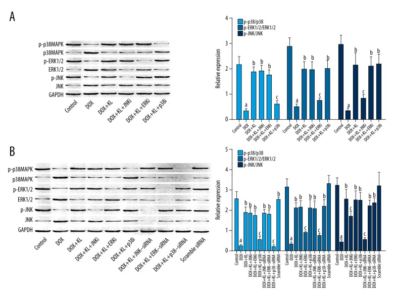
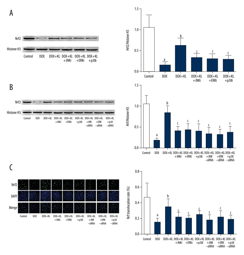
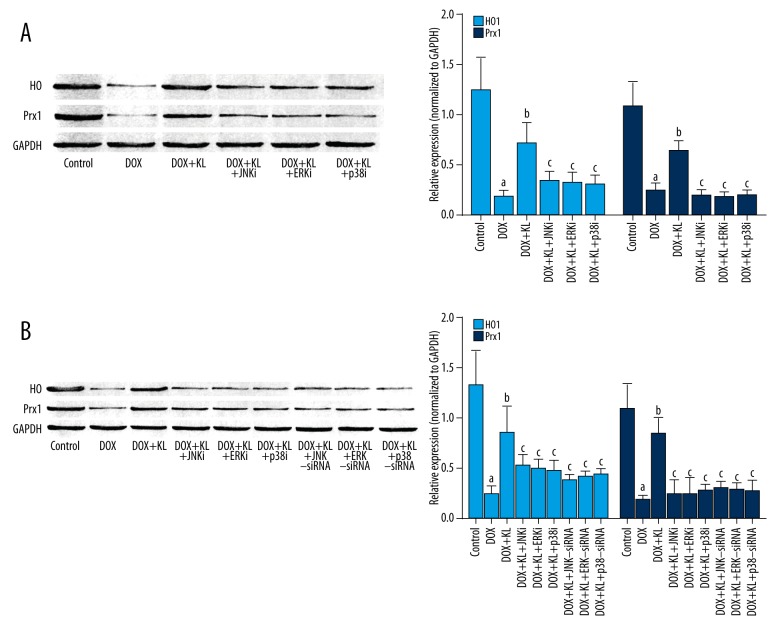
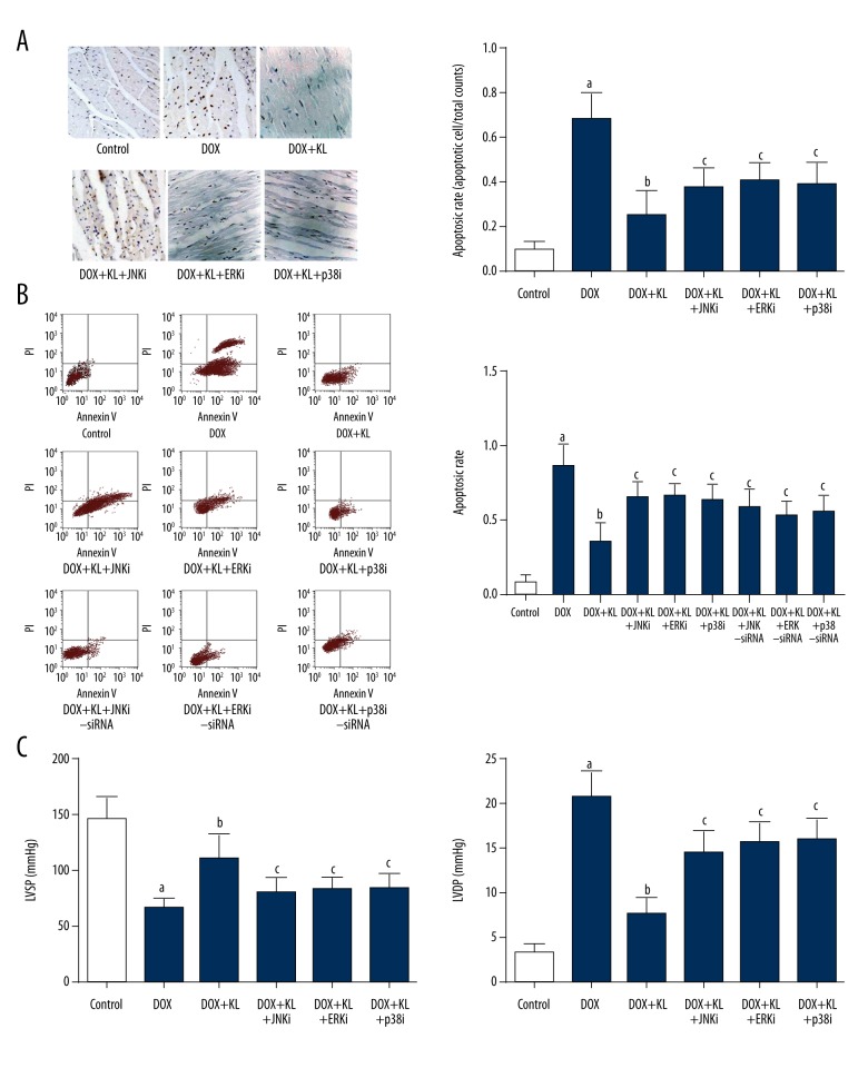
BACKGROUND Anthracyclines-induced cardiotoxicity has become one of the major restrictions of their clinical applications. Klotho showed cardioprotective effects. This study aimed to investigate the effects and possible mechanisms of klotho on doxorubicin (DOX)-induced cardiotoxicity. MATERIAL AND METHODS Rats and isolated myocytes were exposed to DOX and treated with exogenous klotho. Specific inhibitors and siRNAs silencing MAPKs were also used to treat the animals and/or myocytes. An invasive hemodynamic method was used to determine cardiac functions. Intracellular ROS generation was evaluated by DHE staining. Western blotting was used to determine the phosphorylation levels of JNK, ERK, and p38 MAPKs in plasma extracts and Nrf2 in nuclear extracts. Nuclear translocation of Nrf2 in myocytes was evaluated by immunohistochemistry. Cell apoptosis was evaluated by TUNEL assay and flow cytometry. RESULTS Klotho treatment improved DOX-induced cardiac dysfunction in rats. The DOX-induced ROS accumulation and cardiac apoptosis were attenuated by klotho. Impaired phosphorylations of MAPKs, Nrf2 translocation and expression levels of HO1 and Prx1 were also attenuated by klotho treatment. However, the anti-oxidant and anti-apoptotic effects of klotho on DOX-exposed myocardium and myocytes were impaired by both specific inhibitors and siRNAs against MAPKs. Moreover, the recovery effects of klotho on phosphorylations of MAPKs, Nrf2 translocation and expression levels of HO1 and Prx1 were also impaired by specific inhibitors and siRNAs against MAPKs. CONCLUSIONS By recovering the activation of MAPKs signaling, klotho improved cardiac function loss which was triggered by DOX-induced ROS mediated cardiac apoptosis.


Figures

Figure 1
Figure 1
Klotho reduced DOX-induced ROS in myocardium and myocytes which was reversed by MAPKs inhibitors or siRNAs. (A) Left panel demonstrates the captured fluorescent images of DHE stained myocardium. Intracellular ROS is tagged red. Columns on the right side indicate the mean fluorescence intensities of ROS in myocardium harvested from control, DOX, DOX+KL, DOX+KL+JNKi, DOX+KL+ERKi and DOX+KL+p38i respectively. (B) Images on the left side are DHE staining of cultured myocytes. Columns on the right side indicate the mean fluorescence intensities of ROS in myocardium harvested from control, DOX, DOX+KL, DOX+KL+JNKi, DOX+KL+ERKi, DOX+KL+p38i, DOX+KL+JNK-siRNA, DOX+KL+ERK-siRNA, and DOX+KL+p38-siRNA respectively. a Differences were significant when compared with control (p<0.05); b differences were significant when compared with DOX (p<0.05); c differences were significant when compared with DOX+KL (p<0.05).
Figure 2
Figure 2
Klotho activated MAPKs in DOX-treated myocardium and myocytes which was reversed by MAPKs inhibitors or siRNAs. (A, B) The left panels show the immunoblots of p-p38 MAPK, p38 MAPK, p-ERK1/2, ERK1/2, p-JNK, JNK, and GAPDH in cytoplasmic protein samples extracted from myocardium and myocytes. Columns on the right panels indicate the ratio of p-p38MAPK/p38MAPK (light blue), p-ERK2/1/ERK1/2 (blue), and p-JNK/JNK (dark blue) in myocardium and cultured myocytes respectively. a Differences were significant when compared with control (p<0.05); b differences were significant when compared with DOX (p<0.05); c differences were significant when compared with DOX+KL (p<0.05).
Figure 3
Figure 3
Klotho improved nuclear translocation of Nrf2 in DOX-exposed myocardium and myocytes which was reversed by MAPKs inhibitors or siRNAs. (A, B) The left panels show the immunoblots of Nrf2 and Histone H3 in nuclear protein samples extracted from myocardium and myocytes. Columns on the right panels indicate the ratio of Nrf2/histone H3 in myocardium and cultured myocytes respectively. (C) The left panel demonstrates the captured fluorescent images of Nrf2, DAPI, and their merged images in cultured myocytes. Columns on the right part indicate the Nrf2 nuclear translocation rate in different groups. a Differences were significant when compared with control (p<0.05); b differences were significant when compared with DOX (p<0.05); c differences were significant when compared with DOX+KL (p<0.05).
Figure 4
Figure 4
Klotho increased expression levels of anti-oxidant enzymes in DOX-treated myocardium and myocytes which was reversed by MAPKs inhibitors or siRNAs. (A, B) The immunoblots of HO1, Prx1, and GAPDH in myocardium and cultured myocytes in different groups are shown on the left panels. Columns on the right part indicate the ratio of HO/GAPDH and Prx1/GAPDH in myocardium and cultured myocytes respectively. a Differences were significant when compared with control (p<0.05); b differences were significant when compared with DOX (p<0.05); c differences were significant when compared with DOX+KL (p<0.05).
Figure 5
Figure 5
Klotho attenuated DOX-induced apoptosis and cardiac dysfunction which were reversed by treatment of MAPKs and siRNAs. (A) Left panel demonstrates the captured images of TUNEL assay in myocardium. Apoptotic cells are stained brown. Columns on the right part indicate the cell apoptotic rate in different groups. (B) Charts of flow cytometry of apoptosis in cultured myocytes in different groups are shown on the left side. Columns on the right part indicate the cell apoptotic rate in different groups. (C) Columns in this panel indicate the detected LVSP (left side) and LVDP (right side) by hemodynamic method in rats from different groups. a Differences were significant when compared with control (p<0.05); b differences were significant when compared with DOX (p<0.05); c differences were significant when compared with DOX+KL (p<0.05).

References

    1. Xu F, Li X, Liu L, et al. Attenuation of doxorubicin-induced cardiotoxicity by esculetin through modulation of Bmi-1 expression. Exp Ther Med. 2017;14(3):2216–20. - PMC - PubMed
    1. Johnson R, Shabalala S, Louw J, et al. Aspalathin reverts doxorubicin-induced cardiotoxicity through increased autophagy and decreased expression of p53/mTOR/p62 signaling. Molecules. 2017;22(10) pii: E1589. - PMC - PubMed
    1. Hamirani Y, Fanous I, Kramer CM, et al. Anthracycline- and trastuzumab-induced cardiotoxicity: A retrospective study. Med Oncol. 2016;33(7):82. - PMC - PubMed
    1. Lou Y, Wang Z, Xu Y, et al. Resveratrol prevents doxorubicin-induced cardiotoxicity in H9c2 cells through the inhibition of endoplasmic reticulum stress and the activation of the Sirt1 pathway. Int J Mol Med. 2015;36(3):873–80. - PubMed
    1. de Oliveira BL, Niederer S. A biophysical systems approach to identifying the pathways of acute and chronic doxorubicin mitochondrial cardiotoxicity. PLoS Comput Biol. 2016;12(11):e1005214. - PMC - PubMed

MeSH terms