. 2017 Sep 16;8(45):78289-78299.
doi: 10.18632/oncotarget.20961. eCollection 2017 Oct 3.

MeCP2 mediated dysfunction in senescent EPCs

Affiliations

MeCP2 mediated dysfunction in senescent EPCs

Chunli Wang et al. Oncotarget. .

Abstract

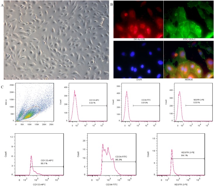
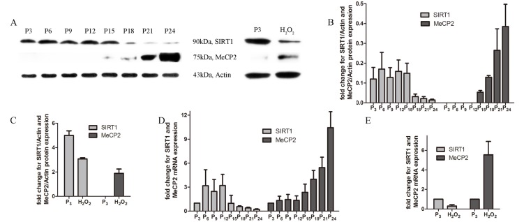
Aging endothelial progenitor cells (EPCs) exhibit functional impairment in terms of proliferation, migration and survival. SIRT1 plays an important role in improving EPCs function. MeCP2, another important epigenetic regulator, is involved in regulating many life-related activities such as cell growth, death and senescence. Here we aim to explore the effect of MeCP2 on the functional activities of senescent EPCs and the underlying mechanisms. By using western blot and real-time PCR, we found that the expression levels of MeCP2 were up-regulated and SIRT1 were down-regulated with replicative senescence and H2O2-induced senescence. Through transduction with adenoviral vectors, EPCs overexpressing MeCP2 had significantly reduced EPCs function, and silencing MeCP2 improved EPCs function. In addition, the protein and mRNA levels of SIRT1 were decreased with MeCP2 overexpression and increased with MeCP2 knockdown. Through co-transfection of EPCs with MeCP2 and SIRT1, we observed that SIRT1 could reverse the effects of MeCP2 on EPCs. In summary, our work demonstrated that MeCP2 inhibited SIRT1 in senescent EPCs.

Keywords: EPCs; Gerotarget; MeCP2; SIRT1; senescence.


Conflict of interest statement

CONFLICTS OF INTEREST There are no conflicts of interest to disclose.

Figures

Figure 1
Figure 1. Cultivation and identification of EPCs derived from umbilical cord blood
A. EPCs exhibited a cobblestone-like cell monolayer at 14 days after seeding(X100). B. Uptake of Dil-Ac-LDL and binding of FITC-UEA-1 were observed with a fluorescence microscope (X400). C. A representative FSC/SSC plot and the expression of EPC markers (CD34, CD133 and VEGFR-2) analyzed by flow cytometry.
Figure 2
Figure 2. Characteristic of the senescence model
A. Cell viability with different concentration of H2O2 determined by CCK-8 assay, EPCs was co-cultured with different concentration of H2O2 for 24 h, then examined the cell viability by CCK-8. B. The morphology of three groups(X100). C. D. Beta-gal staining of the three groups (X100) (the results of positive blue stained cell numbers are reported as mean±SD of three independent experiments, *P<0.05 vs. P3).
Figure 3
Figure 3. Hallmarks of a senescent model
A. Growth curve of the three groups (different groups of EPCs were cultured by six consecutive days of testing using the CCK-8 kit). B. Reactive oxygen assay using DCFH-DA. C. Telomerase activity concentration of the three groups. D. Age-related gene mRNA expression. (data were reported as mean±SD, n=3, *P<0.05 vs. P3).
Figure 4
Figure 4. Function comparison of the three groups
A. Cell cycle by flow cytometry(proliferation index (PI) was used to compare, PI=(S+G2M)/(G0/1+S+G2M)). B. Migration ability by transwell assay(X100, the number of dyed purple cells in three randomly microscopic fields were used to access migration ability). C. Tube formation of the three groups(X40, total loops were used to evaluate the tube formation ability) D. Cell apoptosis by flow cytometry(the percentage of apoptotic cells with Annexin V positive expression was used to assess apoptosis). (three independent experiments were repeated, and the same result were done)
Figure 5
Figure 5. Expression of MeCP2 and SIRT1 with aging
A., B., C. Protein levels of MeCP2 and SIRT1 by western blot with replicative senescence and H2O2-induced senescence. D., E. mRNA levels of MeCP2 and SIRT1 by RT-PCR with replicative senescence and H2O2-induced senescence (experiments were repeated three times, and the same trend resulted).
Figure 6
Figure 6. EPCs transfected with Ad-MeCP2 or Ad-sh-MeCP2
A. Fluorescence images of EPCs transfected with Ad-MeCP2 or Ad-sh-MeCP2 at different MOIs. B., C., E., F. The effects of Ad-MeCP2 (MOI 100) and Ad-sh-MeCP2 (MOI 100) on the expression of MeCP2 were confirmed by western blot. D., G. The effects of Ad-MeCP2 (MOI 100) and Ad-sh-MeCP2 (MOI 100) on the expression of MeCP2 were confirmed by RT-PCR analysis. H., I. SIRT1 expression of EPCs transfected with Ad-MeCP2 or Ad-sh-MeCP2 was examined by western blot. J. SIRT1 expression of EPCs transfected with Ad-MeCP2 or Ad-sh-MeCP2 was examined by RT-PCR analysis. (n=3, *P<0.05 vs. GFP).
Figure 7
Figure 7. Functional alterations with overexpression or silencing by co-transfection of MeCP2 and SIRT1
A. Proliferation by flow cytometry (proliferation index (PI) was used to compare the proliferation ability, PI=(S+G2M)/(G0/1+S+G2M)). B. Migration ability by Transwell assay (X100, the number of dyed purple cells in three randomly microscopic fields were used to access migration ability). C. Tube formation of EPCs (X40, total loops were used to evaluate the tube formation ability). (three independent experiments were repeated, and the same result were done).

References

    1. Altabas V, Altabas K, Kirigin L. Endothelial progenitor cells (EPCs) in ageing and age-related diseases: How currently available treatment modalities affect EPC biology, atherosclerosis, and cardiovascular outcomes. Mech Ageing Dev. 2016;159:49–62. - PubMed
    1. Rauscher FM, Goldschmidt-Clermont PJ, Davis BH, Wang T, Gregg D, Ramaswami P, Pippen AM, Annex BH, Dong C, Taylor DA. Aging, progenitor cell exhaustion, and atherosclerosis. Circulation. 2003;108:457–463. - PubMed
    1. Simper D, Wang S, Deb A, Holmes D, McGregor C, Frantz R, Kushwaha SS, Caplice NM. Endothelial progenitor cells are decreased in blood of cardiac allograft patients with vasculopathy and endothelial cells of noncardiac origin are enriched in transplant atherosclerosis. Circulation. 2003;108:143–149. - PubMed
    1. Xu Q. Mouse models of arteriosclerosis: from arterial injuries to vascular grafts. Am J Pathol. 2004;165:1–10. - PMC - PubMed
    1. Lee SH, Lee JH, Yoo SY, Hur J, Kim HS, Kwon SM. Hypoxia inhibits cellular senescence to restore the therapeutic potential of old human endothelial progenitor cells via the hypoxia-inducible factor-1alpha-TWIST-p21 axis. Arterioscler Thromb Vasc Biol. 2013;33:2407–2414. - PubMed

LinkOut - more resources