

Metapristone (RU486 metabolite) suppresses NSCLC by targeting EGFR-mediated PI3K/AKT pathway
- PMID: 29108234
- PMCID: PMC5667967
- DOI: 10.18632/oncotarget.18640
Metapristone (RU486 metabolite) suppresses NSCLC by targeting EGFR-mediated PI3K/AKT pathway
Abstract
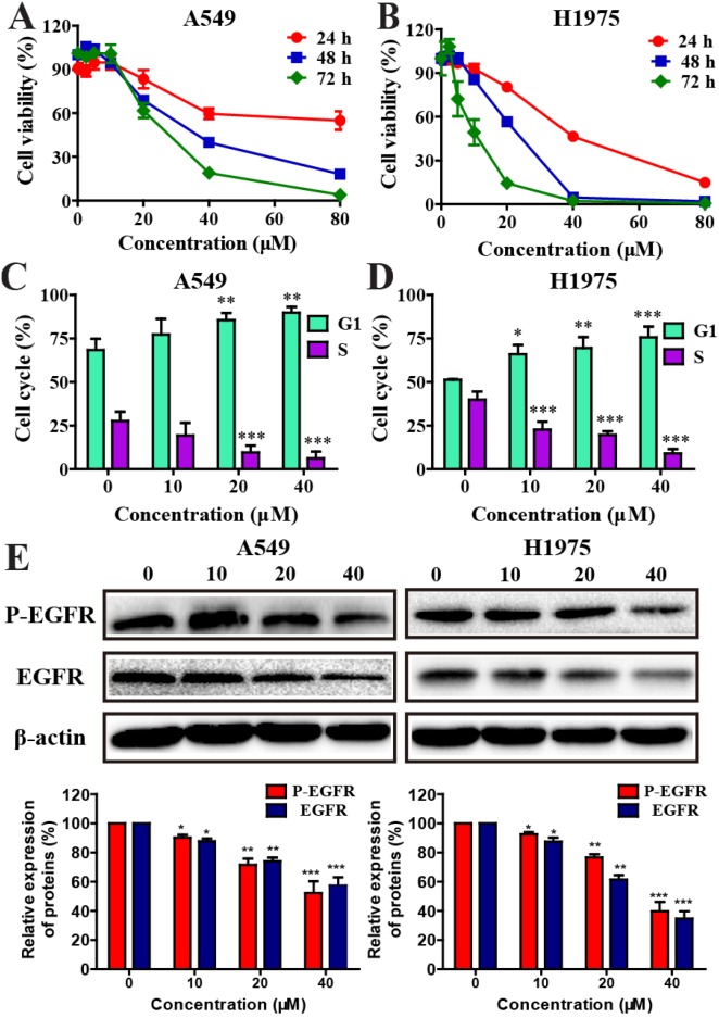
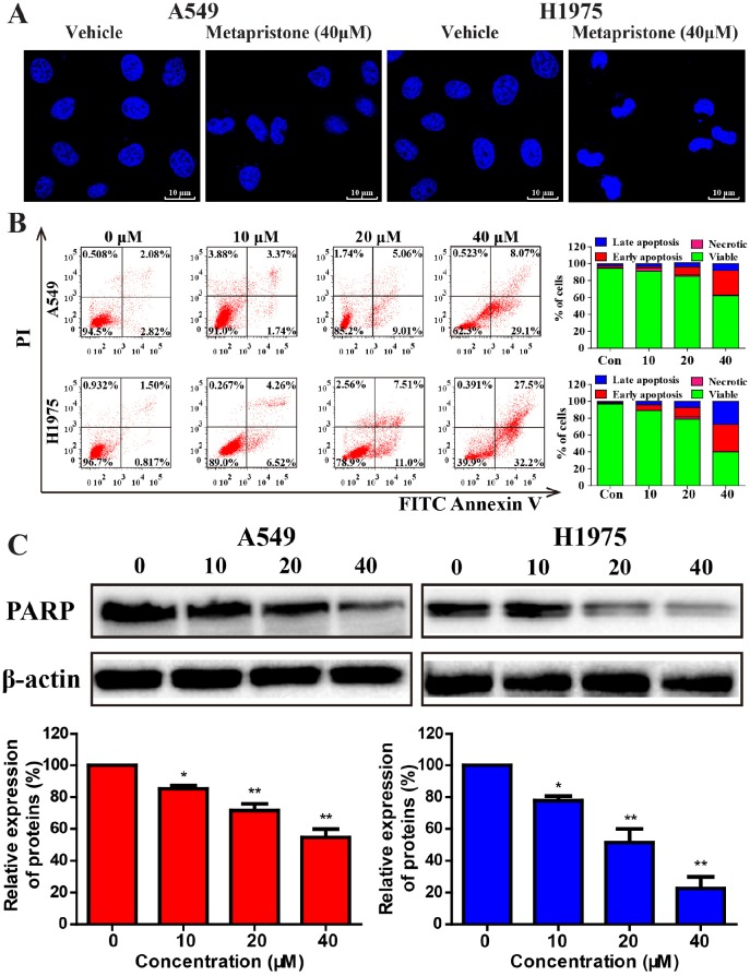
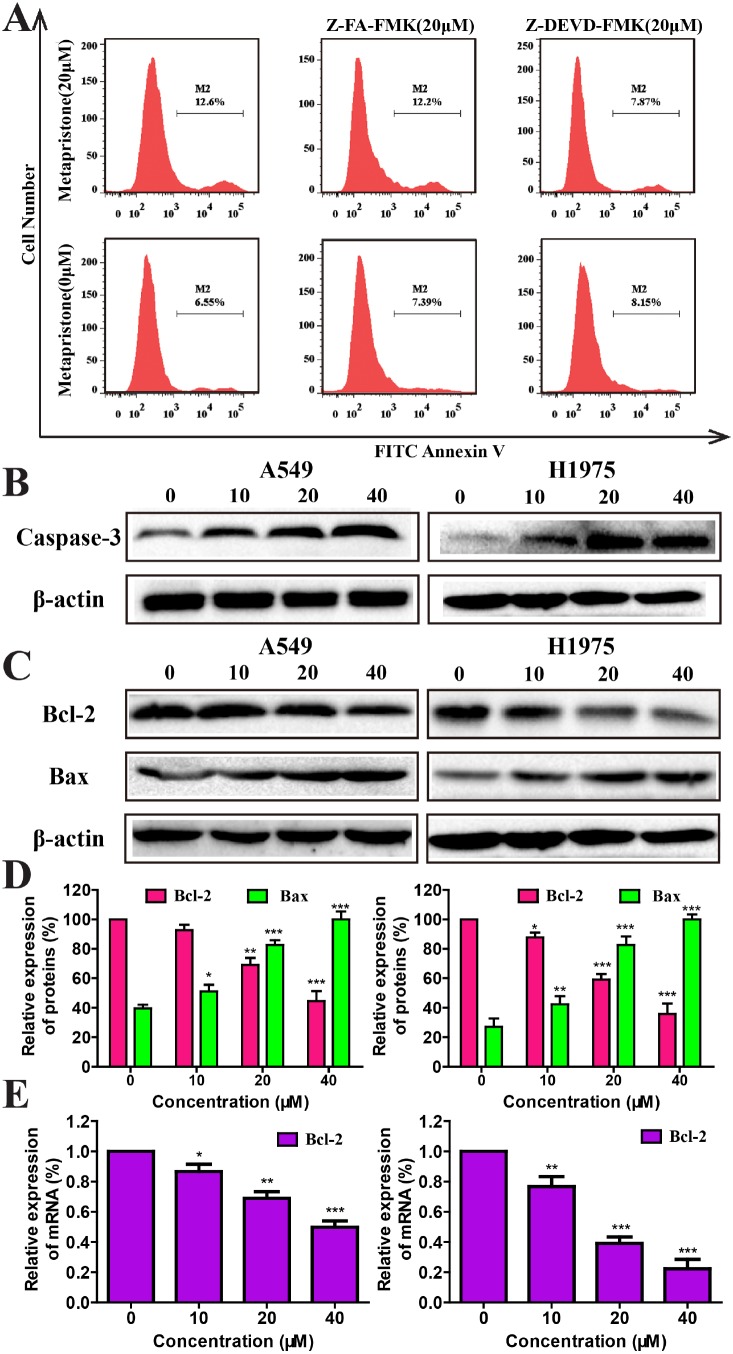
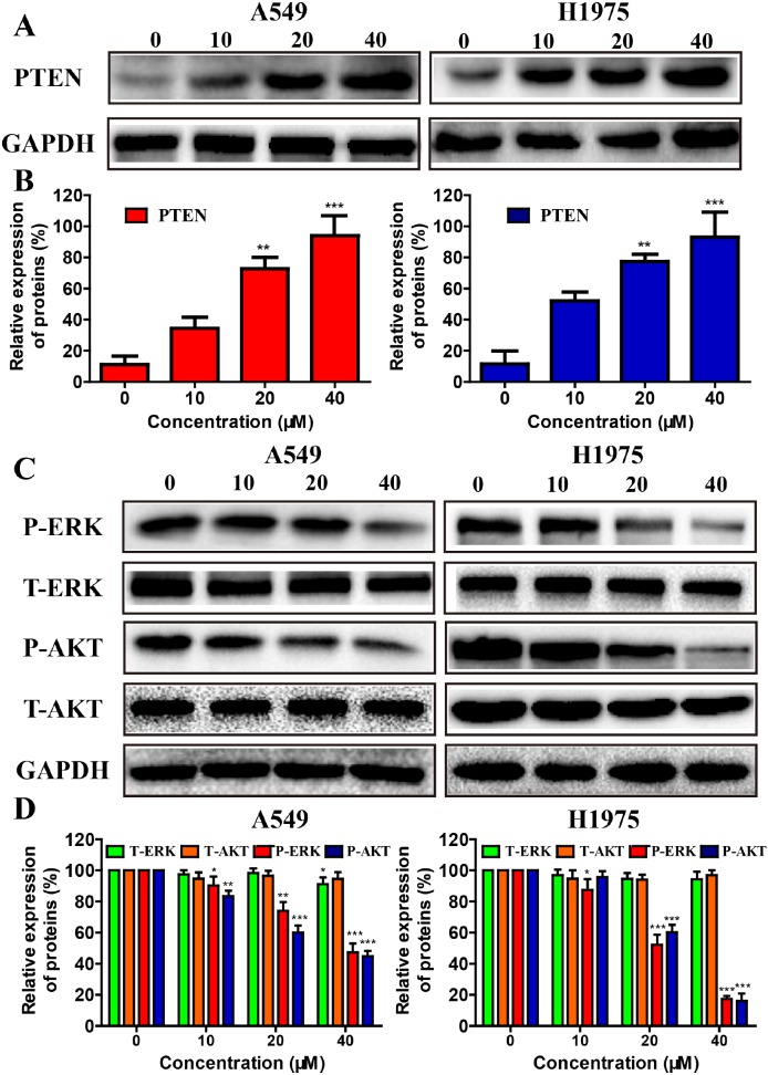
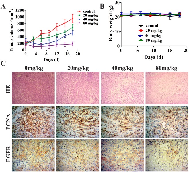
Therapies targeting epidermal growth factor receptor (EGFR) can effectively treat with non-small cell lung cancer (NSCLC), but NSCLC's drug resistance makes it intractable. Herein, we showed that RU486 metabolite metapristone inhibited the proliferation of various NSCLC cell lines with either wild (A549, H1299, H520) or mutated EGFR (H1975, HCC827). The suppression was resulted from inhibition by metapristone of EGFR signaling pathways through down-regulating the EGFR, PTEN, as well as AKT and ERK proteins. In addition, metapristone inhibited anti-apoptotic marker Bcl-2, and activated pro-apoptotic key signaling proteins caspase-3, and poly (ADP-ribose) polymerase. Metapristone induced A549 and H1975 cell cycle via arrest at the G0-G1 stage. What's more, metapristone inhibited the growth of NSCLC xenografts in BALB/c nude mice through decreasing the expression of tumor growth biomarkers PCNA and EGFR. Taken together, the present study demonstrated that metapristone suppressed NSCLC proliferation by promoting apoptosis via decrease the cellular EGFR-mediated PI3K/AKT pathways. The results suggest metapristone a new treatment for EGFR-overexpressed NSCLC.
Keywords: EGFR; cell apoptosis; cell proliferation; metapristone; non-small cell lung cancer.
Conflict of interest statement
CONFLICTS OF INTEREST Thes authors declare no competing financial interest.
Figures

Similar articles
-
Co-delivery of sorafenib and metapristone encapsulated by CXCR4-targeted PLGA-PEG nanoparticles overcomes hepatocellular carcinoma resistance to sorafenib.J Exp Clin Cancer Res. 2019 May 31;38(1):232. doi: 10.1186/s13046-019-1216-x. J Exp Clin Cancer Res. 2019. PMID: 31151472 Free PMC article.
-
Metapristone suppresses non-small cell lung cancer proliferation and metastasis via modulating RAS/RAF/MEK/MAPK signaling pathway.Biomed Pharmacother. 2017 Jun;90:437-445. doi: 10.1016/j.biopha.2017.03.091. Epub 2017 Apr 6. Biomed Pharmacother. 2017. PMID: 28391165
-
Metapristone (RU486 derivative) inhibits cell proliferation and migration as melanoma metastatic chemopreventive agent.Biomed Pharmacother. 2017 Jun;90:339-349. doi: 10.1016/j.biopha.2017.03.076. Epub 2017 Apr 2. Biomed Pharmacother. 2017. PMID: 28376402
-
PA-MSHA in combination with EGFR tyrosine kinase inhibitor: A new strategy to overcome the drug resistance of non-small cell lung cancer cells.Oncotarget. 2016 Aug 2;7(31):49384-49396. doi: 10.18632/oncotarget.9891. Oncotarget. 2016. PMID: 27283902 Free PMC article.
-
Delphinidin reduces cell proliferation and induces apoptosis of non-small-cell lung cancer cells by targeting EGFR/VEGFR2 signaling pathways.PLoS One. 2013 Oct 4;8(10):e77270. doi: 10.1371/journal.pone.0077270. eCollection 2013. PLoS One. 2013. PMID: 24124611 Free PMC article.
Cited by
-
Ergosterol peroxide from marine fungus Phoma sp. induces ROS-dependent apoptosis and autophagy in human lung adenocarcinoma cells.Sci Rep. 2018 Dec 18;8(1):17956. doi: 10.1038/s41598-018-36411-2. Sci Rep. 2018. PMID: 30560887 Free PMC article.
-
Co-delivery of sorafenib and metapristone encapsulated by CXCR4-targeted PLGA-PEG nanoparticles overcomes hepatocellular carcinoma resistance to sorafenib.J Exp Clin Cancer Res. 2019 May 31;38(1):232. doi: 10.1186/s13046-019-1216-x. J Exp Clin Cancer Res. 2019. PMID: 31151472 Free PMC article.
-
Interleukin-22 enhances chemoresistance of lung adenocarcinoma cells to paclitaxel.Hum Cell. 2020 Jul;33(3):850-858. doi: 10.1007/s13577-020-00373-3. Epub 2020 May 25. Hum Cell. 2020. PMID: 32452013
-
Metapristone (RU486-derivative) inhibits endometrial cancer cell progress through regulating miR-492/Klf5/Nrf1 axis.Cancer Cell Int. 2021 Jan 7;21(1):29. doi: 10.1186/s12935-020-01682-1. Cancer Cell Int. 2021. PMID: 33413440 Free PMC article.
-
Effect of Mifepristone on Migration and Proliferation of Oral Cancer Cells.Int J Mol Sci. 2024 Aug 12;25(16):8777. doi: 10.3390/ijms25168777. Int J Mol Sci. 2024. PMID: 39201464 Free PMC article.
References
-
- Torre LA, Siegel RL, Jemal A. Lung cancer statistics. Adv Exp Med Biol. 2016;893:1–19. - PubMed
-
- Torre LA, Bray F, Siegel RL, Ferlay J, Lortet-Tieulent J, Jemal A. Global cancer statistics, 2012. CA Cancer J Clin. 2015;65:87–108. - PubMed
-
- Hirsch FR, Scagliotti GV, Mulshine JL, Kwon R, Curran WJ, Jr, Wu YL, Paz-Ares L. Lung cancer: current therapies and new targeted treatments. Lancet. 2017;389:299–311. - PubMed
-
- Gridelli C, Rossi A, Carbone DP, Guarize J, Karachaliou N, Mok T, Petrella F, Spaggiari L, Rosell R. Non-small-cell lung cancer. Nat Rev Dis Primers. 2015;1:15009. - PubMed
LinkOut - more resources
Full Text Sources
Other Literature Sources
Research Materials
Miscellaneous
