Stress During Development of Experimental Endometriosis Influences Nerve Growth and Disease Progression
- PMID: 29108503
- PMCID: PMC6343219
- DOI: 10.1177/1933719117737846
Stress During Development of Experimental Endometriosis Influences Nerve Growth and Disease Progression
Abstract
Purpose: We have previously shown that stress prior to induction worsens clinical presentation and inflammatory parameters in a rat model of endometriosis. This study was designed to examine whether stress during the development of endometriosis can affect the growth of endometriotic implants through nerve growth and immune alterations.
Methods: Endometriosis was surgically induced in female Sprague-Dawley rats by suturing uterine horn implants onto the small intestine mesentery. Two weeks later, one group of rats (endo-stress) was subjected to a 10-day swim stress protocol. Controls had no stress (endo-no stress) or sutures only and stress (sham-stress). On day 60, all rats were killed and examined for the presence of endometriotic vesicles. The size of each vesicle was measured. The uterus and colon were removed and assessed for damage, cell infiltration, and expression of nerve growth factor (NGF), its receptors (p75 and Tropomyosin receptor kinase A (Trk-A)/pTrk-A), and calcitonin gene-related peptide, a sensory fiber marker. A differential analysis of peritoneal fluid white blood cell count was performed.
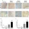
Results: Stress significantly increased endometriotic vesicle size but not colonic damage and increased infiltration of mast cells. Significantly increased expression of NGF and its receptors was found in the uterus of animals with endometriosis receiving stress.
Conclusions: Stress stimulates the development of ectopic endometrial vesicles in an animal model of endometriosis and increases inflammatory cell recruitment to the peritoneum. In addition, stress promotes nerve fiber growth in the uterus.
Keywords: Trk; endometriosis; nerve growth factor; p75; stress.
Conflict of interest statement
Figures


Similar articles
-
Stress exacerbates endometriosis manifestations and inflammatory parameters in an animal model.Reprod Sci. 2012 Aug;19(8):851-62. doi: 10.1177/1933719112438443. Epub 2012 Apr 23. Reprod Sci. 2012. PMID: 22527982 Free PMC article.
-
Nerve Growth Factor Is Associated With Sexual Pain in Women With Endometriosis.Reprod Sci. 2018 Apr;25(4):540-549. doi: 10.1177/1933719117716778. Epub 2017 Jul 4. Reprod Sci. 2018. PMID: 28673205
-
Endometriosis as a neurovascular condition: estrous variations in innervation, vascularization, and growth factor content of ectopic endometrial cysts in the rat.Am J Physiol Regul Integr Comp Physiol. 2008 Jan;294(1):R162-71. doi: 10.1152/ajpregu.00649.2007. Epub 2007 Oct 17. Am J Physiol Regul Integr Comp Physiol. 2008. PMID: 17942489 Free PMC article.
-
The role of nerve growth factor and its receptors in tumorigenesis and cancer pain.Biosci Trends. 2014 Apr;8(2):68-74. doi: 10.5582/bst.8.68. Biosci Trends. 2014. PMID: 24815383 Review.
-
Manipulation of the nerve growth factor network in prostate cancer.Expert Opin Investig Drugs. 2007 Mar;16(3):303-9. doi: 10.1517/13543784.16.3.303. Expert Opin Investig Drugs. 2007. PMID: 17302525 Review.
Cited by
-
Endometriosis and pain in the adolescent- striking early to limit suffering: A narrative review.Neurosci Biobehav Rev. 2020 Jan;108:866-876. doi: 10.1016/j.neubiorev.2019.12.004. Epub 2019 Dec 17. Neurosci Biobehav Rev. 2020. PMID: 31862211 Free PMC article. Review.
-
Voluntary Wheel Running Reduces Vesicle Development in an Endometriosis Animal Model Through Modulation of Immune Parameters.Front Reprod Health. 2021;3:826541. doi: 10.3389/frph.2021.826541. Front Reprod Health. 2021. PMID: 36284640 Free PMC article.
-
Suppressive regulatory T cells and latent transforming growth factor-β-expressing macrophages are altered in the peritoneal fluid of patients with endometriosis.Reprod Biol Endocrinol. 2018 Feb 1;16(1):9. doi: 10.1186/s12958-018-0325-2. Reprod Biol Endocrinol. 2018. PMID: 29391020 Free PMC article.
-
WERF Endometriosis Phenome and Biobanking Harmonisation Project for Experimental Models in Endometriosis Research (EPHect-EM-Homologous): homologous rodent models.Mol Hum Reprod. 2025 Jul 3;31(3):gaaf021. doi: 10.1093/molehr/gaaf021. Mol Hum Reprod. 2025. PMID: 40628400 Free PMC article.
-
The COVID-19 pandemic and patients with endometriosis: A survey-based study conducted in Turkey.Int J Gynaecol Obstet. 2020 Nov;151(2):249-252. doi: 10.1002/ijgo.13339. Epub 2020 Sep 1. Int J Gynaecol Obstet. 2020. PMID: 32750726 Free PMC article.
References
-
- Tariverdian N, Rucke M, Szekeres-Bartho J, et al. Neuroendocrine circuitry and endometriosis: progesterone derivative dampens corticotropin-releasing hormone-induced inflammation by peritoneal cells in vitro. J Mol Med. 2010;88(3):267–278. - PubMed
-
- Toth B. Stress, inflammation and endometriosis: are patients stuck between a rock and a hard place? J Mol Med. 2010;88(3):223–225. - PubMed
-
- Huntington A, Gilmour JA. A life shaped by pain: women and endometriosis. J Clin Nurs. 2005;14(9):1124–1132. - PubMed
-
- Barnack JL, Chrisler JC. The experience of chronic illness in women: a comparison between women with endometriosis and women with chronic migraine headaches. Women Health. 2007;46(1):115–133. - PubMed
Publication types
MeSH terms
Substances
Grants and funding
LinkOut - more resources
Full Text Sources
Other Literature Sources
Medical
Research Materials

