Review
. 2018 Mar;1862(3):532-546.
doi: 10.1016/j.bbagen.2017.11.005. Epub 2017 Nov 3.

Matrix vesicles from chondrocytes and osteoblasts: Their biogenesis, properties, functions and biomimetic models

Affiliations
Review

Matrix vesicles from chondrocytes and osteoblasts: Their biogenesis, properties, functions and biomimetic models

Massimo Bottini et al. Biochim Biophys Acta Gen Subj. 2018 Mar.

Abstract

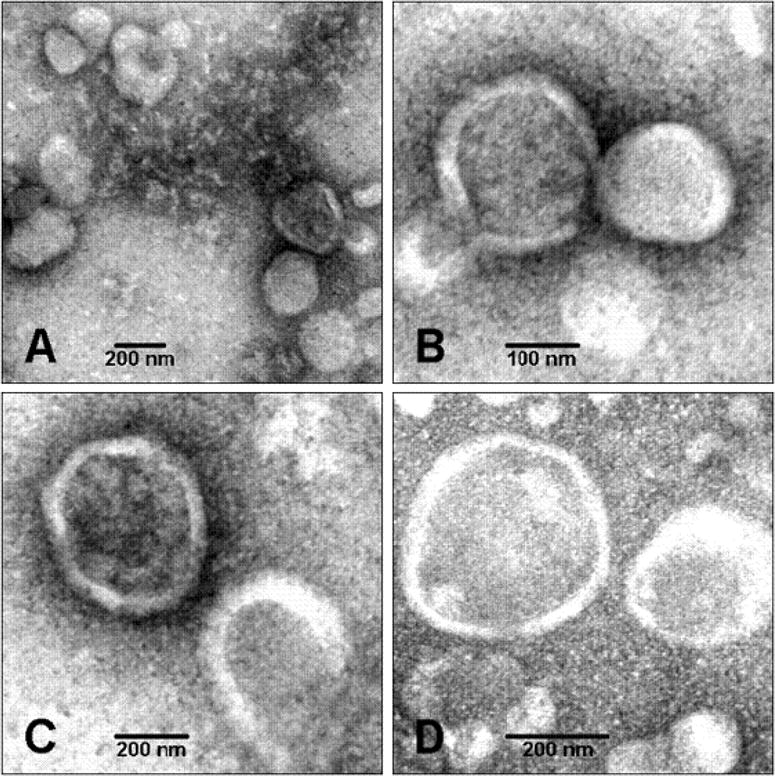
Background: Matrix vesicles (MVs) are released from hypertrophic chondrocytes and from mature osteoblasts, the cells responsible for endochondral and membranous ossification. Under pathological conditions, they can also be released from cells of non-skeletal tissues such as vascular smooth muscle cells. MVs are extracellular vesicles of approximately 100-300nm diameter harboring the biochemical machinery needed to induce mineralization.

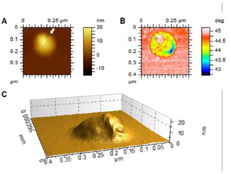
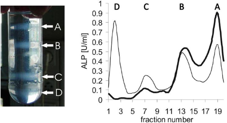
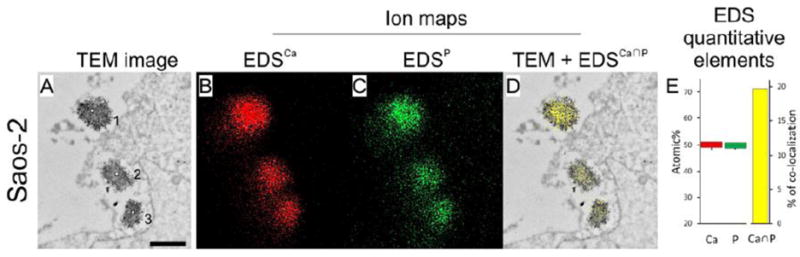
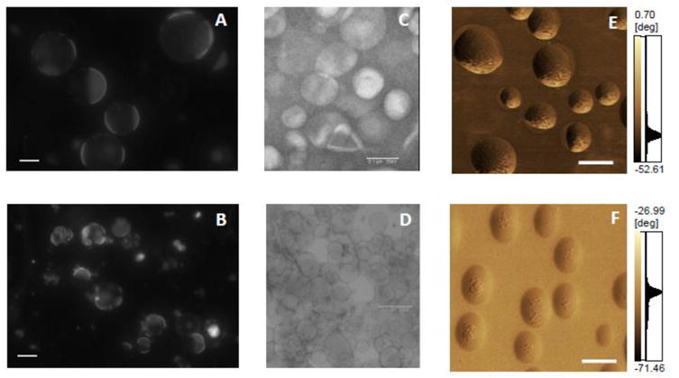
Scope of the review: The review comprehensively delineates our current knowledge of MV biology and highlights open questions aiming to stimulate further research. The review is constructed as a series of questions addressing issues of MVs ranging from their biogenesis and functions, to biomimetic models. It critically evaluates experimental data including their isolation and characterization methods, like lipidomics, proteomics, transmission electron microscopy, atomic force microscopy and proteoliposome models mimicking MVs.

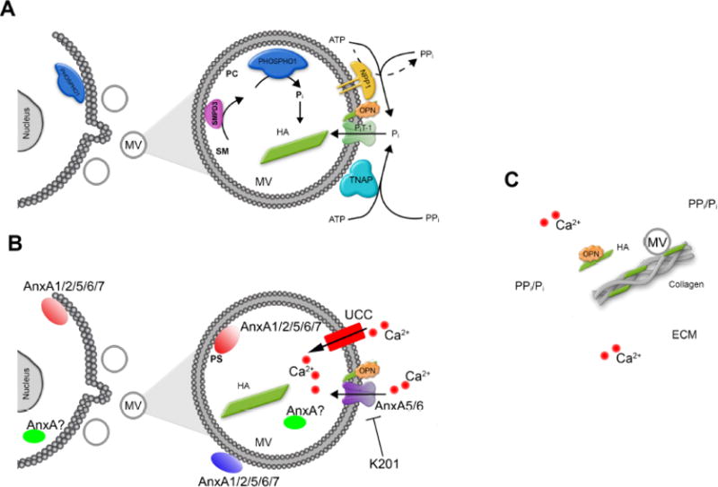
Major conclusions: MVs have a relatively well-defined function as initiators of mineralization. They bind to collagen and their composition reflects the composition of lipid rafts. We call attention to the as yet unclear mechanisms leading to the biogenesis of MVs, and how minerals form and when they are formed. We discuss the prospects of employing upcoming experimental models to deepen our understanding of MV-mediated mineralization and mineralization disorders such as the use of reconstituted lipid vesicles, proteoliposomes and, native sample preparations and high-resolution technologies.

General significance: MVs have been extensively investigated owing to their roles in skeletal and ectopic mineralization. MVs serve as a model system for lipid raft structures, and for the mechanisms of genesis and release of extracellular vesicles.

Keywords: Atomic force microscopy; Electron microscopy; Lipid raft; Matrix vesicles; Mineralization; Proteoliposomes.


Figures

Figure 1
Figure 1. Electron transmission micrographs of MVs
MVs extracted from chicken embryo growth plate cartilages were round structures with a diameter ranging from 100 to 250 nm. (Magnifications: (A), × 53,000; (B), × 100,000; (C), × 75,000; (D), × 100,000). Adapted from [20].
Figure 2
Figure 2. Atomic force microscope image of an MV released by primary chondrocytes
Cells were isolated from WT mouse and grown in differentiation medium (α-MEM with 10% FBS and 50 mg/mL ascorbic acid) for 18 days. MVs were isolated through digestion of the cell monolayer with collagenase followed by two-step differential ultracentrifugation, and dropped onto a freshly cleaved mica substrate. The substrate was dried under vacuum and scanned by means of an AFM (model 5500 AFM, Keysight Technologies, Santa Rosa, CA). (A), topography image; (B), phase image; (C), three-dimensional reconstruction of the topography image along the direction indicated by the white arrow in A.
Figure 3
Figure 3. Schematic depiction of our current understanding of the biochemical pathways involved in MV-mediated initiation of skeletal, dental and vascular mineralization
For the sake of simplicity, the main functional components in MVs are divided in two parts: (A), Pi turnover. Currently available data are compatible with the following sequence of events: MVs initiate mineral deposition by accumulation of Pi generated intravesicularly by the action of PHOSPHO1 on phosphocholine (PC) derived from sphingomyelin (SM) by the action of SMPD3, and also via PiT-1-mediated incorporation of Pi generated extravesicularly by TNAP and/or NPP1. The extravesicular propagation of mineral onto the collagenous matrix is mainly controlled by the pyrophosphatase activity of TNAP that restricts the concentration of PPi, a potent mineralization inhibitor, to establish a PPi/Pi ratio conducive to controlled mineralization. How MVs are formed is still unclear but emerging evidence indicates that PHOSPHO1 is involved in MV biogenesis. (B), Ca2+ turnover. Mineral deposition in MVs is initiated by accumulation of Ca2+ intravesicularly by the action of calcium carriers like annexins (AnxA), or unidentified calcium carriers (UCC). Annexins may be located in the lumen of MV or may bind to phosphoserine (PS)-rich membrane domains on the inner surface, or the outer surface of MVs. Some annexins at slightly intracellular acidic pH may protrude MV membrane and form transmembrane like ion channels. Benzodiazepine derivative K201 was known to be a potential inhibitor of annexin calcium channel activity and reduced the ability of the MVs to subsequently mineralize collagen fibers. (C), Propagation of apatite crystals in the ECM. It is unclear how apatite crystals formed within MVs propagate onto the collagenous matrix. The attachment of MV to the collagenous extracellular matrix (ECM) via a number of collagen-binding proteins has been proposed. Osteopontin (OPN), or fetuin A (not shown), which are potent mineralization inhibitors that bind to apatite as soon as it is exposed to the extracellular fluid, further restricts the degree of ECM mineralization.
Figure 4
Figure 4. Membrane fraction profiles of collagenase-released MVs or of MV enriched microsomes without collagenase treatment
Femurs from 17-old day chicken embryo were washed in synthetic cartilage lymph medium. They were homogenized and subjected with or without collagenase treatments. Then, membrane fractions were isolated during several differential ultracentrifugations. The pellet was subjected by a sucrose gradient composed (from the bottom to the top) of 1.6 M (3 ml), 1.2 M (8 ml), 0.9 M (9 ml), and 0.6 M (9 ml) sucrose dissolved in synthetic cartilage lymph. Then it was centrifuged at 100,000×g for 60 min at 4°C. Left panel: MV enriched microsomes obtained without collagenase treatment indicated four membranous fractions (A-D) after ultracentrifugation. Right panel: Collagenase-treatment released two membrane fractions (A, B, bold line). One fraction (A) corresponds to MVs as indicated by high alkaline phosphatase activity (ALP). Omission of collagenase treatment leaded to four membrane fractions (A, B, C, D, normal line) having less MVs as indicated by lower ALP activity in fraction A. Fraction A was unambiguously assigned to MVs due to its mineralization property. Adapted from [45].
Figure 5
Figure 5. Transmission electron microscopy coupled with X-ray microanalysis of osteoblast like Saos-2 cells
Saos-2 osteosarcoma cells were stimulated for mineralization by treatment with 50 μg mL−1 ascorbic acid and 7.5 mM beta-glycerophosphate for 7 days. Cells were washed in physiological desensitization medium, fixed with paraformaldehyde/glutaraldehyde mixture for 1h and postfixed with osmium tetroxide for 20 min. After dehydration in ethanol series probes were embedded in LR White resin at 56°C for 48 hrs. Finally the samples were cut with ultramicrotome for 700Å sections and counterstained with uranyl acetate for 1h followed by lead citrate for 2 min. The cells with vesicles were observed under high performance TEM JEM-1400 (JEOL Co., Japan) with a magnification of x50,000 (A, bar 500 nm) and elemental maps (B, Calcium, red and C, Phosphorus, green) were performed using energy-dispersive full range X-ray microanalysis system EDS INCA ENERGY TEM (Oxford Instruments, UK). Presence of calcium and phosphoreus were evidenced in vesicles containing dense materials. Co-localization of both elements (D, yellow) and quantitative determinations (E, Calcium, red, Phosphorus, green, Calcium to Phosphorus, yellow), provided evidence of calcium and phosphorus deposition inside vesicles, suggesting apatite deposition in MVs. Adapted from [149].
Figure 6
Figure 6. Characterization of liposomes and proteoliposomes by different techniques
Fluorescence microscopy (60× magnification, scales bar of 20 μm, labeled with 2% rhodamine) of (A) giant liposomes consisting of DOPC, CHOL and SM (8:1:1, molar ratio) and (B) 8:1:1 DOPC:CHOL:SM-giant liposomes harboring TNAP. Electron microscopy using negative staining of: (C) Liposomes consisting of DPPC (D) DPPC-proteoliposomes harboring TNAP, both with 50× magnification. 3D topographic AFM images of (E) liposomes consisting of DPPC and DPPS (9:1, molar ratio) (xy area is 1 μm × 11 μm and z axis from 0 to 84.50 nm) and scale bar of 250 nm and (F) 9:1 DPPC:DPPS-proteoliposomes harboring TNAP (xy area is 2.50 μm × 2.50 μm and z axis from 0 to 40.81 nm) and scale bar of 250 nm.

References

    1. Anderson HC. Matrix vesicles and calcification. Curr Rheumatol Rep. 2003;5:222–226. - PubMed
    1. Anderson HC, Mulhall D, Garimella R. Role of extracellular membrane vesicles in the pathogenesis of various diseases, including cancer, renal diseases, atherosclerosis, and arthritis. Lab Invest. 2010;90:1549–1557. - PubMed
    1. Goldberg M, Kulkarni AB, Young M, Boskey A. Dentin: structure, composition and mineralization. Front Biosci (Elite Ed) 2011;3:711–35. - PMC - PubMed
    1. Stratmann U, Schaarschmidt K, Wiesmann HP, Plate U, Höhling HJ. Mineralization during matrix-vesicle-mediated mantle dentine formation in molars of albino rats: a microanalytical and ultrastructural study. Cell Tissue Res. 1996;284:223–30. - PubMed
    1. Shanahan CM, Crouthamel MH, Kapustin A, Giachelli CM. Arterial calcification in chronic kidney disease: key roles for calcium and phosphate. Circ Res. 2011;109:697–711. - PMC - PubMed

Publication types

MeSH terms