. 2018 Feb;20(2):81-88.
doi: 10.1016/j.micinf.2017.10.008. Epub 2017 Nov 8.

Effect of Mycobacterium tuberculosis infection on adipocyte physiology

Affiliations

Effect of Mycobacterium tuberculosis infection on adipocyte physiology

Janeesh Plakkal Ayyappan et al. Microbes Infect. 2018 Feb.

Abstract

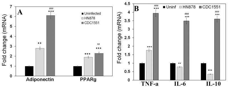
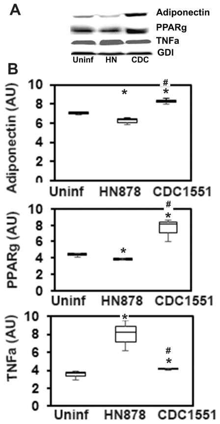
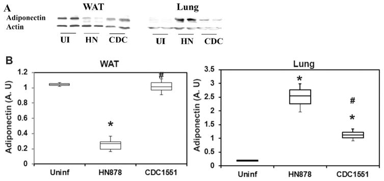
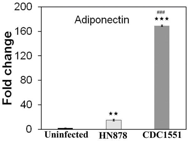
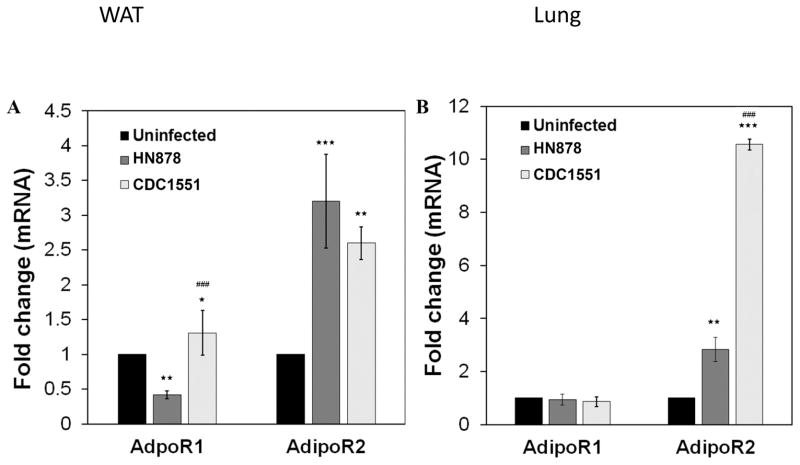
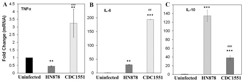
Tuberculosis (TB) remains as a major threat to human health worldwide despite of the availability of standardized antibiotic therapy. One of the characteristic of pathogenic Mycobacterium tuberculosis (Mtb), the causative agent of tuberculosis is its ability to persist in the host in a dormant state and develop latent infection without clinical signs of active disease. However, the mechanisms involved in bacterial persistence and the establishment of latency is not well understood. Adipose tissue is emerging as an important niche that favors actively replicating as well as dormant Mtb during acute and latent infection. This also suggests that Mtb can disseminate from the lungs to adipose tissue during aerosol infection and/or from adipose tissue to lungs during reactivation of latent infection. In this study, we report the interplay between key adipokine levels and the dynamics of Mtb pathogenesis in the lungs and adipose tissue using a rabbit model of pulmonary infection with two clinical isolates that produce divergent outcome in disease progression. Results show that markers of adipocyte physiology and function were significantly altered during Mtb infection and distinct patterns of adipokine expression were noted between adipose tissue and the lungs. Moreover, these markers were differentially expressed between active disease and latent infection. Thus, this study highlights the importance of targeting adipocyte function as potential target for developing better TB intervention strategies.

Keywords: Adiponectin; Adipose tissue; Inflammation; Latency; Tuberculosis.


Conflict of interest statement

6. CONFLICT OF INTEREST STATEMENT

None of the authors have conflict of interest.

Figures

Figure 1
Figure 1. qPCR analysis demonstrated different pattern of adipokines mRNA expression levels in the WAT of rabbits aerosol infected with HN878 (active TB) and CDC1551 (latent TB)
The fold changes in the mRNA levels were calculated after comparing to control rabbits and normalizing to housekeeping HPRT mRNA levels (n=4, 12 weeks post infection). (A) Fold change in the levels of adiponectin and PPARγ. (B) Fold change in the levels of adipocytokines TNFα, IL-6 and IL-10. Significance represent mean values of the data with Standard Error of the mean (SEM) as vertical lines. (The error bars represent standard error of the mean. ** p ≤ 0.01 or *** p ≤ 0.001 compared to uninfected rabbits; ##p ≤ 0.01 or ###p ≤ 0.001 compared to HN878 infected rabbits).
Figure 2
Figure 2. Immunoblot analysis demonstrated a significant alteration in the levels of adiponectin, PPARγ and TNFα in 3T3-l1 adipocytes infected with different strains of Mtb (MOI 3:1) for 48h
(A) Protein analysis of adipogenic factors (adiponectin and PPARγ) and proinflammatory TNFα levels in the adipocytes infected with Mtb strains HN878 or CDC1551 compared to uninfected cells. (B) Band intensity was measured using Image J software and normalized to GDI, and represented as Box-Whisker plots (n=3, experiment was duplicated). (The error bars represent standard error of the mean. *p ≤ 0.05 compared to uninfected rabbits; #p ≤ 0.05 compared to HN878 infected rabbits).
Figure 3
Figure 3. Immunoblot analysis of adiponectin in WAT and lungs during Mtb infection in rabbit models
(A) Protein levels of adiponectin in Wat and Lungs of rabbits infected with Mtb strains HN878 or CDC1551 compared to uninfected rabbits. (B) Band intensity was measured using Image J software and normalized to GDI, and represented as Box-Whisker plots. (n=3) (The error bars represent standard error of the mean. * p ≤ 0.05 or $p ≤ 0.5 compared to uninfected rabbits; #p ≤ 0.05 compared to HN878 infected rabbits).
Figure 4
Figure 4. qPCR analysis demonstrated increased adiponectin mRNA expression in the lungs during Mtb infection in rabbit models infected with HN878 (active TB) and CDC1551 (latent TB) Mtb strains
The fold change in the mRNA levels was calculated after comparing to control rabbits and normalizing to housekeeping HPRT mRNA levels. (n=4, 12 weeks post infection. The error bars represent standard error of the mean. ** p ≤ 0.01 or *** p ≤ 0.001 compared to uninfected rabbits; ##p ≤ 0.01 or ###p ≤ 0.001 compared to HN878 infected rabbits).
Figure 5
Figure 5. qPCR analysis demonstrated different pattern of adiponectin receptors R1 and R2 mRNA expression levels in the WAT (A) and lungs (B) of rabbits aerosol infected with HN878 (active TB) and CDC1551 (latent TB)
The fold changes in the mRNA levels were calculated after comparing to control rabbits and normalizing to housekeeping HPRT mRNA levels (n=4, 12 weeks post infection). (The error bars represent standard error of the mean. * p≤ 0.05, ** p ≤ 0.01 or *** p ≤ 0.001 compared to uninfected rabbits; ##p ≤ 0.01 or ###p ≤ 0.001 compared to HN878 infected rabbits).
Figure 6
Figure 6. qPCR analysis demonstrated different pattern of inflammatory cytokines mRNA expression levels in the lungs of rabbits aerosol infected with HN878 (active TB) and CDC1551 (latent TB)
The fold changes in the mRNA levels of pro-inflammatory cytokines (A) TNFα and (B) IL-6 and anti-inflammatory cytokine (C) IL-10 in the lungs of rabbits infected with HN878 or CDC1551 compared to uninfected. The fold changes were calculated after comparing to control rabbits and normalizing to housekeeping HPRT mRNA levels (n=4, 12 weeks post infection). (The error bars represent standard error of the mean. ** p ≤ 0.01 or *** p ≤ 0.001 compared to uninfected rabbits; ##p ≤ 0.01 or ###p ≤ 0.001 compared to HN878 infected rabbits).

References

    1. Global tuberculosis report. http://www.who.int/tb/publications/global_report/gtbr15_main_text.pdf.
    1. http://www.who.int/tb/publications/global_report/gtbr2016_main_text.pdf?ua=1

    1. Selwyn PA, Hartel D, Wasserman W, Drucker E. Impact of the AIDS epidemic on morbidity and mortality among intravenous drug users in a New York City methadone maintenance program. Am J Public Health. 1989;79:1358–62. - PMC - PubMed
    1. Oni T, Stoever K, Wilkinson RJ. Tuberculosis, HIV, and type 2 diabetes mellitus: a neglected priority. Lancet Respir Med. 2013;1:356–58. - PubMed
    1. Guariguata L, Whiting DR, Hambleton I, Beagley J, Linnenkamp U, Shaw JE. Global estimates of diabetes prevalence for 2013 and projections for 2035. Diabetes Res Clin Pract. 2014;103:137–49. - PubMed

Publication types

MeSH terms