Review
. 2017 Dec 22;292(51):21170-21179.
doi: 10.1074/jbc.R117.807560. Epub 2017 Nov 6.

Peptide splicing by the proteasome

Affiliations
Review

Peptide splicing by the proteasome

Nathalie Vigneron et al. J Biol Chem. .

Abstract

The proteasome is the major protease responsible for the production of antigenic peptides recognized by CD8+ cytolytic T cells (CTL). These peptides, generally 8-10 amino acids long, are presented at the cell surface by major histocompatibility complex (MHC) class I molecules. Originally, these peptides were believed to be solely derived from linear fragments of proteins, but this concept was challenged several years ago by the isolation of anti-tumor CTL that recognized spliced peptides, i.e. peptides composed of fragments distant in the parental protein. The splicing process was shown to occur in the proteasome through a transpeptidation reaction involving an acyl-enzyme intermediate. Here, we review the steps that led to the discovery of spliced peptides as well as the recent advances that uncover the unexpected importance of spliced peptides in the composition of the MHC class I repertoire.

Keywords: MHC class I; antigen presentation; antigen processing; cytolytic T lymphocytes; major histocompatibility complex (MHC); peptides; proteasome; spliced peptides.


Conflict of interest statement

The authors declare that they have no conflicts of interest with the contents of this article.

Figures
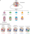
Figure 1.
Figure 1.
Structure and composition of proteasomes. A, structure of the proteasome and its regulators. The 20S proteasome is a cylindrical particle composed of four stacked heptameric rings (α1–7–β1–7–β1–7–α1–7) that delineate a chamber in which the proteins are degraded. The two outer rings are made of α subunits (beige), and the two inner rings are made of β subunits (red/bluish gray). Three types of β subunits are catalytically active (red). The α rings form the gates of the proteasome that control the entry of intracellular proteins inside the proteasome chamber. They can interact with different types of regulatory particles as follows: 19S regulatory particles (PA700), PA28αβ, PA28γ, PA200, and PI31. These regulatory particles can bind to one or both sides of the 20S particle or form hybrid proteasomes where the 20S core binds two different regulators. B, proteasome subtypes. Depicted are the five proteasome subtypes, which differ in their catalytic subunits. The standard proteasome contains the constitutive catalytic subunits β1, β2, and β5, whereas the immunoproteasome contains the interferon-γ-inducible catalytic subunits β1i, β2i, and β5i. The intermediate proteasomes express a mixed assortment of constitutive and inducible subunits; the intermediate proteasome β5i contains β1, β2, and β5i and the intermediate proteasome β1i β5i contains β1i, β2, and β5i. Finally, the thymoproteasome contains catalytic subunit β5t, which is homologous to β5 and β5i and is specifically expressed in the cortical thymic epithelial cells along with β1i and β2i.
Figure 2.
Figure 2.
Proteasome catalytic activities. A, hydrolytic activity of the proteasome. The hydroxyl group of the N-terminal threonine of the catalytic subunits of the proteasome attacks the carbonyl group of the peptide bond. This leads to the production of an acyl-enzyme intermediate, in which a peptide fragment remains attached to the proteasome by an ester link. Then a water molecule hydrolyzes the ester link between the peptide and the threonine residue, thereby restoring the hydroxyl group of the catalytic threonine and producing the C-terminal end of the antigenic peptide. B, peptide splicing occurs by transpeptidation. Various synthetic peptides were combined in a pairwise manner and incubated with 20S proteasomes. The presence of the peptide RTK_QLYPEW in the digests was determined using either a specific CTL or by mass spectrometry. The presence (+) or absence (−) of the peptide in the digest is indicated. Ac-QLYPEW, N-α-acetylated peptide QLYPEW. C, peptide splicing in the proteasome. Here, the splicing of the antigenic peptide RTK_QLYPEW derived from the gp100 is shown. Following formation of the acyl-enzyme intermediate involving the fragment RTK, the free N-terminal amino group of peptide QLYPEW present in the proteasome chamber attacks the acyl-enzyme intermediate leading to the formation of the peptide RTK_QLYPEW composed of two peptide fragments originally distant in the protein. D, reverse splicing reaction in the proteasome. The splicing of the antigenic peptide SLPRGT_STPK derived from the SP110 is shown. Following formation of the acyl-enzyme intermediate involving the fragment SLPRGT, the free N-terminal amino group of peptide STPK attacks the acyl-enzyme intermediate leading to the formation of the peptide SLPRGT_STPK. Because in the protein SP110, the C-terminal reactant STPK is located upstream from the N-terminal reactant SLPRGT, the final peptide is composed of two peptide fragments originally distant in the protein and spliced together in reverse order to that in which they occur in the protein.
Figure 3.
Figure 3.
Lack of physiological relevance of trans-splicing. Shown is an experiment designed to visualize the occurrence of splicing of fragments originating from two different proteins (i.e. trans-splicing). COS-7 cells were transfected with pairs of constructs mutated at critical residues so that the only way to produce the antigenic peptide was to splice fragments originating from two different proteins. The amount of IFNγ produced by the CTL after incubation with the transfected cells is shown. The results indicate that trans-splicing does not occur at a significant level in cells.
Figure 4.
Figure 4.
Production of the tyrosinase peptide IYMDGTADFSF. Upon entry into the ER (step 1), tyrosinase is glycosylated at specific asparagine residues (step 2). Some of the glycosylated tyrosinase protein is then retrotranslocated from the ER into the cytosol, likely due to misfolding (step 3). Upon deglycosylation by PNGase, asparagines are deamidated into aspartate residues (step 4). The aspartate-containing tyrosinase is then degraded by the proteasome (step 5), a process that leads to the reverse splicing of two fragments, each containing a post-translationally modified aspartate.
Figure 5.
Figure 5.
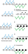
Schematic representation of the splicing reaction occurring at the catalytic site of the proteasome. The peptide to be cleaved is represented by a necklace with pearls indicating individual amino acids. The non-primed binding site is indicated in blue and the primed binding site is in green. Each binding site is subdivided in different pockets. Three pockets are labeled S1 to S3 for the non-primed site and S1′ to S3′ for the primed binding site. Amino acids binding to the non-primed binding site are labeled P1 to Pn, and those binding to the primed binding site are labeled P1′ to Pn′. The peptide splicing reaction starts with the formation of an acyl-enzyme intermediate with the peptide located at the non-primed binding site. In the course of the peptide splicing reaction, the C-terminal splice reactant (black) comes into the vicinity of the acyl-enzyme intermediate, most likely by binding to the primed binding site. An alternative pocket that could fit the C-terminal reactant has been postulated but is not represented here. The amine group of the C-terminal splice reactant then produces a nucleophilic attack on the ester bond of the acyl-enzyme intermediate to create the spliced peptide, which then exits the catalytic site.

References

    1. Rock K. L., Gramm C., Rothstein L., Clark K., Stein R., Dick L., Hwang D., and Goldberg A. L. (1994) Inhibitors of the proteasome block the degradation of most cell proteins and the generation of peptides presented on MHC class I molecules. Cell 78, 761–771 - PubMed
    1. Androlewicz M. J., Anderson K. S., and Cresswell P. (1993) Evidence that transporters associated with antigen processing translocate a major histocompatibility complex class I-binding peptide into the endoplasmic reticulum in an ATP-dependent manner. Proc. Natl. Acad. Sci. U.S.A. 90, 9130–9134 - PMC - PubMed
    1. Saric T., Chang S. C., Hattori A., York I. A., Markant S., Rock K. L., Tsujimoto M., and Goldberg A. L. (2002) An IFN-γ-induced aminopeptidase in the ER, ERAP1, trims precursors to MHC class I-presented peptides. Nat. Immunol. 3, 1169–1176 - PubMed
    1. Saveanu L., Carroll O., Lindo V., Del Val M., Lopez D., Lepelletier Y., Greer F., Schomburg L., Fruci D., Niedermann G., and van Endert P. M. (2005) Concerted peptide trimming by human ERAP1 and ERAP2 aminopeptidase complexes in the endoplasmic reticulum. Nat. Immunol. 6, 689–697 - PubMed
    1. York I. A., Chang S. C., Saric T., Keys J. A., Favreau J. M., Goldberg A. L., and Rock K. L. (2002) The ER aminopeptidase ERAP1 enhances or limits antigen presentation by trimming epitopes to 8–9 residues. Nat. Immunol. 3, 1177–1184 - PubMed

Publication types

MeSH terms

Substances

LinkOut - more resources