. 2018 Jan 22;86(2):e00645-17.
doi: 10.1128/IAI.00645-17. Print 2018 Feb.

Candidalysin Drives Epithelial Signaling, Neutrophil Recruitment, and Immunopathology at the Vaginal Mucosa

Affiliations

Candidalysin Drives Epithelial Signaling, Neutrophil Recruitment, and Immunopathology at the Vaginal Mucosa

Jonathan P Richardson et al. Infect Immun. .

Abstract

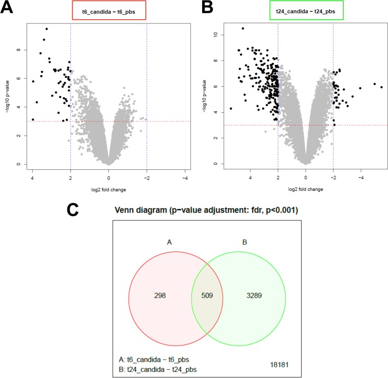
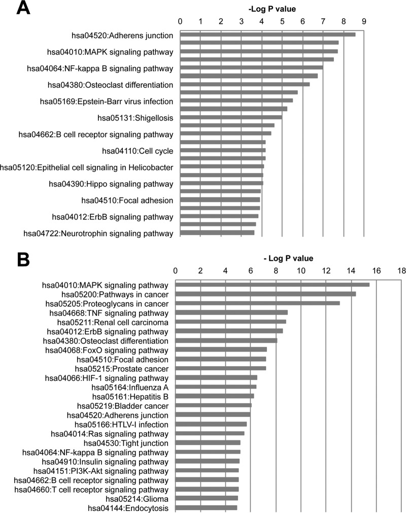
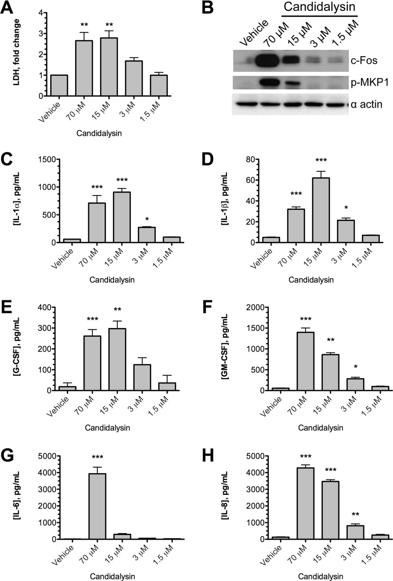
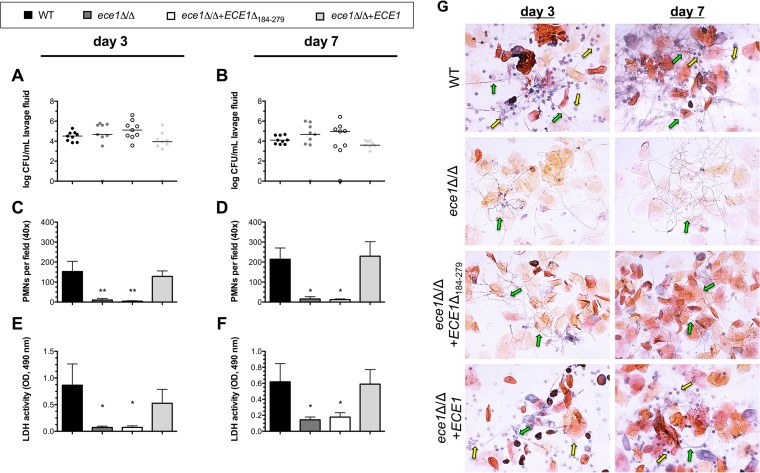
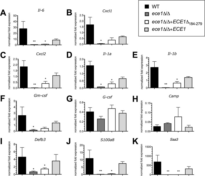
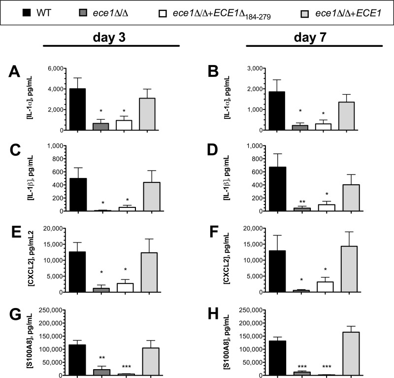
Unlike other forms of candidiasis, vulvovaginal candidiasis, caused primarily by the fungal pathogen Candida albicans, is a disease of immunocompetent and otherwise healthy women. Despite its prevalence, the fungal factors responsible for initiating symptomatic infection remain poorly understood. One of the hallmarks of vaginal candidiasis is the robust recruitment of neutrophils to the site of infection, which seemingly do not clear the fungus, but rather exacerbate disease symptomatology. Candidalysin, a newly discovered peptide toxin secreted by C. albicans hyphae during invasion, drives epithelial damage, immune activation, and phagocyte attraction. Therefore, we hypothesized that Candidalysin is crucial for vulvovaginal candidiasis immunopathology. Anti-Candida immune responses are anatomical-site specific, as effective gastrointestinal, oral, and vaginal immunities are uniquely compartmentalized. Thus, we aimed to identify the immunopathologic role of Candidalysin and downstream signaling events at the vaginal mucosa. Microarray analysis of C. albicans-infected human vaginal epithelium in vitro revealed signaling pathways involved in epithelial damage responses, barrier repair, and leukocyte activation. Moreover, treatment of A431 vaginal epithelial cells with Candidalysin induced dose-dependent proinflammatory cytokine responses (including interleukin 1α [IL-1α], IL-1β, and IL-8), damage, and activation of c-Fos and mitogen-activated protein kinase (MAPK) signaling, consistent with fungal challenge. Mice intravaginally challenged with C. albicans strains deficient in Candidalysin exhibited no differences in colonization compared to isogenic controls. However, significant decreases in neutrophil recruitment, damage, and proinflammatory cytokine expression were observed with these strains. Our findings demonstrate that Candidalysin is a key hypha-associated virulence determinant responsible for the immunopathogenesis of C. albicans vaginitis.

Keywords: Candida; Candidalysin; epithelial cells; immunopathogenesis; mucosal immunity; mucosal pathogens; mycology; vaginitis; vulvovaginal.


Figures

FIG 1
FIG 1
Global differential gene expression induced during C. albicans infection of RVE. Three independent RVE were challenged with C. albicans or mock infected with PBS for 6 or 24 h, and differential gene expression (≥2-fold change) was assessed by microarray analysis. Shown are volcano plots depicting log2-fold expression changes (blue dotted lines) at P < 0.001 (red dotted lines) of genes between the 6-h (A) and 24-h (B) time points following challenge with C. albicans or the PBS mock control. (C) Venn diagram depicting the absolute number of genes expressed between C. albicans at 6 h (red) and 24 h (green), after adjusting for P value and FDR (P < 0.01).
FIG 2
FIG 2
Host signaling pathways predicted to be activated during C. albicans infection of RVE. Based on differential gene expression (≥2-fold change), KEGG pathway analysis using the DAVID Web-based package revealed several host pathways predicted to be significantly (P < 0.001) activated at 6 h (A) or 24 h (B). Pathways are listed in order of highest probability of activation. Significance was assessed using the DAVID statistical package via analysis of variance (ANOVA).
FIG 3
FIG 3
Candidalysin is sufficient to induce cellular damage and proinflammatory responses in vaginal epithelial cells. (A) A431 vaginal epithelial cells were exposed to Candidalysin (1.5, 3, 15, and 70 μM) for 24 h, and cellular damage was quantified by LDH assay. The data are presented as the fold change relative to vehicle control. Statistics were applied relative to the vehicle control (n = 3 biological repeats). (B) Western blot analysis of the vaginal epithelial responses to different concentrations of Candidalysin. Epithelial cell lysates (20 μg total protein) were probed with anti c-Fos and anti p-MKP1 antibodies. One representative blot is presented (from 3 biological repeats). (C to H) Quantification of cytokines (IL-1α, IL-1β, G-CSF, GM-CSF, IL-6, and IL-8) secreted from vaginal epithelial cells in response to different concentrations of Candidalysin. Statistics were applied relative to the vehicle control (n = 3 biological repeats). The graphs are plotted as means plus SEM. (A and C to H) Statistical significance was calculated using one-way ANOVA and Dunnet's posttest. ***, P < 0.001; **, P < 0.01, *, P < 0.05.
FIG 4
FIG 4
Candidalysin is required for neutrophil recruitment and mucosal damage in a murine model of vulvovaginal candidiasis. (A to F) Groups of estrogen-treated C57BL/6 mice (n = 4) were intravaginally challenged with WT, ece1Δ/Δ, ece1Δ/Δ + ECE1Δ184–279, and ece1Δ/Δ + ECE1 strains of C. albicans, and vaginal lavage fluid was assessed longitudinally at day 3 and day 7 for fungal burden (horizontal lines, medians) (A and B), PMNs (means plus SEM) (C and D), or the damage biomarker LDH (means plus SEM) (E and F). Statistical significance was calculated using one-way ANOVA and Tukey's posttest. **, P < 0.01, *, P < 0.05. (G) Papanicolaou staining was performed on smears made from vaginal lavage fluid to assess PMN influx (yellow arrows) and hypha formation (green arrows) at day 3 and day 7 p.i. Representative images are shown. All inoculation groups were performed in duplicate, and the data were combined.
FIG 5
FIG 5
Candidalysin is required for proinflammatory cytokine expression in the murine vagina. Groups of estrogen-treated C57BL/6 mice (n = 4) were intravaginally challenged with WT, ece1Δ/Δ, ece1Δ/Δ + ECE1Δ184–279, and ece1Δ/Δ + ECE1 strains of C. albicans; whole vaginal tissue was excised at day 3 p.i.; and extracted RNA was processed for qPCR analysis. The genes chosen for qPCR were those previously identified as being induced by Candidalysin or C. albicans during in vitro or in vivo challenge: Il-6 (A), Cxcl1 (B), Cxcl2 (C), Il-1a (D), Il-1b (E), Gm-csf (F), G-csf (G), Camp (H), Defb3 (I), S100A8 (J), and Saa3 (K). All the genes were internally compared to the Actb housekeeping gene and to mock-infected controls using the ΔΔCT method. The graphs are plotted as the mean normalized fold expression plus SEM. Statistical significance was calculated using one-way ANOVA and Tukey's posttest. **, P < 0.01, *, P < 0.05.
FIG 6
FIG 6
Candidalysin is required for release of hallmark proinflammatory cytokines and chemokines into the vaginal lavage fluid during murine vaginitis. Groups of estrogen-treated C57BL/6 mice (n = 4) were intravaginally challenged with WT, ece1Δ/Δ, ece1Δ/Δ + ECE1Δ184–279, and ece1Δ/Δ + ECE1 strains of C. albicans, and vaginal lavage fluid was assessed longitudinally by ELISA at day 3 and day 7 p.i. for inflammatory markers: IL-1α (A and B), IL-1β (C and D), Cxcl2 (E and F), and S100A8 (G and H). All inoculation groups were performed in duplicate, and the data were combined. The graphs are plotted as means plus SEM. Statistical significance was calculated using one-way ANOVA and Tukey's posttest. ***, P < 0.001; **, P < 0.01, *, P < 0.05.

References

    1. Achkar JM, Fries BC. 2010. Candida infections of the genitourinary tract. Clin Microbiol Rev 23:253–273. doi:10.1128/CMR.00076-09. - DOI - PMC - PubMed
    1. Sobel JD. 2007. Vulvovaginal candidosis. Lancet 369:1961–1971. doi:10.1016/S0140-6736(07)60917-9. - DOI - PubMed
    1. Sobel JD. 1997. Vaginitis. N Engl J Med 337:1896–1903. doi:10.1056/NEJM199712253372607. - DOI - PubMed
    1. Fidel PL Jr, Barousse M, Espinosa T, Ficarra M, Sturtevant J, Martin DH, Quayle AJ, Dunlap K. 2004. An intravaginal live Candida challenge in humans leads to new hypotheses for the immunopathogenesis of vulvovaginal candidiasis. Infect Immun 72:2939–2946. doi:10.1128/IAI.72.5.2939-2946.2004. - DOI - PMC - PubMed
    1. Peters BM, Yano J, Noverr MC, Fidel PL Jr. 2014. Candida vaginitis: when opportunism knocks, the host responds. PLoS Pathog 10:e1003965. doi:10.1371/journal.ppat.1003965. - DOI - PMC - PubMed

Publication types

MeSH terms