. 2017 Nov 21;114(47):E10216-E10223.
doi: 10.1073/pnas.1713897114. Epub 2017 Nov 6.

Definition of the hypothalamic GnRH pulse generator in mice

Affiliations

Definition of the hypothalamic GnRH pulse generator in mice

Jenny Clarkson et al. Proc Natl Acad Sci U S A. .

Abstract

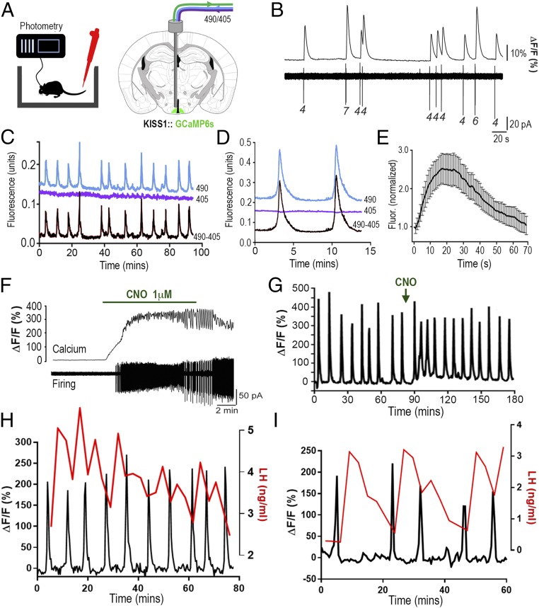
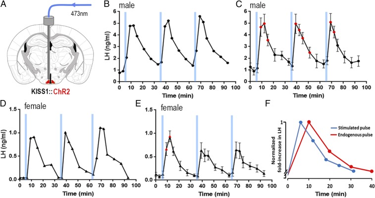
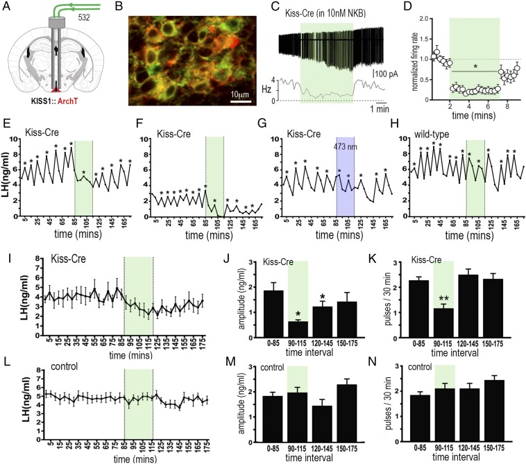
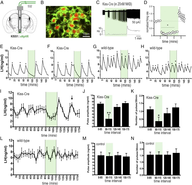
The pulsatile release of luteinizing hormone (LH) is critical for mammalian fertility. However, despite several decades of investigation, the identity of the neuronal network generating pulsatile reproductive hormone secretion remains unproven. We use here a variety of optogenetic approaches in freely behaving mice to evaluate the role of the arcuate nucleus kisspeptin (ARNKISS) neurons in LH pulse generation. Using GCaMP6 fiber photometry, we find that the ARNKISS neuron population exhibits brief (∼1 min) synchronized episodes of calcium activity occurring as frequently as every 9 min in gonadectomized mice. These ARNKISS population events were found to be near-perfectly correlated with pulsatile LH secretion. The selective optogenetic activation of ARNKISS neurons for 1 min generated pulses of LH in freely behaving mice, whereas inhibition with archaerhodopsin for 30 min suppressed LH pulsatility. Experiments aimed at resetting the activity of the ARNKISS neuron population with halorhodopsin were found to reset ongoing LH pulsatility. These observations indicate the ARNKISS neurons as the long-elusive hypothalamic pulse generator driving fertility.

Keywords: GnRH; arcuate nucleus; fertility; kisspeptin; pulse.


Conflict of interest statement

The authors declare no conflict of interest.

Figures

Fig. 1.
Fig. 1.
ARNKISS neurons exhibit periodic synchronizations in intracellular calcium concentrations tightly correlated with pulsatile LH secretion in gonadectomized male mice. (A) KISS1-Cre mice injected with Cre-dependent GCaMP6s AAV into the ARN (green) and connected to a fiber photometry system with regular blood sampling from the tail tip. Alternating 490-nm (Ca2+-dependent) and 405-nm (Ca2+-independent) wavelength light passes down the photometry fiber. (B) Simultaneous GCaMP6s calcium (Top) and cell-attached (Bottom) recordings from an ARNKISS neuron. Numbers indicate spikes per burst. (C) Fluorescence emission traces (490-nm calcium, blue; 405-nm background, purple) recorded from ARNKISS neurons of a gonadectomized male mouse with the subtracted signal (490 − 405) shown below. (D) Continuous recording of two calcium events from a gonadectomized mouse. (E) Average fluorescence waveform of calcium events (16 events, 7 mice) normalized to the beginning of each event. (F) Simultaneous brain slice recording of GCaMP6s fluorescence (Top) and action potential firing (Bottom) from a GCaMP6s/hM3Dq-expressing ARNKISS neuron showing the response to CNO applied in the bath. (G) Representative trace showing the effect of i.p. CNO (arrow) on GCaMP6 calcium signal from ARNKISS neurons in a gonadectomized mouse. (H and I) Representative dual calcium and plasma LH traces showing the near-perfect correlation of ARNKISS neuron GCaMP6 calcium events (black) with pulses of LH secretion (red) in two separate gonadectomized male mice. (I) The mouse with the slowest calcium events is shown.
Fig. 2.
Fig. 2.
Brief 1-min channelrhodopsin activation of ARNKISS neurons evokes pulsatile LH secretion in freely behaving intact male and female mice. (A) Schematic of experiment. (B) Repeated 1-min blue light (473-nm) activations of ARNKISS neurons evoke LH pulses in an intact male mouse. (C) Mean (±SEM) LH levels (red symbols indicate significantly elevated LH levels; repeated-measures ANOVA with Dunnett’s post hoc tests; n = 5). (D) Repeated 1-min blue light (473-nm) activations of ARNKISS neurons evoke LH pulses in an intact female mouse. (E) Mean (±SEM) LH levels (red symbols indicate significantly elevated LH levels; repeated-measures ANOVA with Dunnett’s post hoc tests; n = 6). (F) Overlaid profiles of endogenous (red) and ChR2-activated (blue) LH pulses in intact male mice. Pulses are normalized for fold increase over baseline; endogenous LH pulses (n = 6) were sampled every 10 min and ChR2-activated LH pulses (n = 15) every 6 min.
Fig. 3.
Fig. 3.
Archaerhodopsin inhibition of ARNKISS neurons suppresses pulsatile LH secretion in gonadectomized female mice. (A) KISS1-Cre mice injected with Cre-dependent ArchT-tdTomato AAV into the ARN (red) and implanted with bilateral optic fibers in the ARN. (B) Dual fluorescence image of ARNKISS neurons expressing ArchT (red) and kisspeptin (GFP reporter). (C) Action potential firing in an ArchT-expressing ARNKISS neuron in the presence of 10 nM NKB in a brain slice. The neuron responds to green light illumination (shading) with a decrease in firing followed by a return to control levels. (C, Top) Action potential firing. (C, Bottom) Rate meter for the same cell. (D) Mean (±SEM) normalized firing rate of ARNKISS neurons (n = 11) responding to green light. *P < 0.05 compared with baseline, Friedman test. (EH) Pulsatile LH secretion in ArchT KISS1-Cre (EG) and wild-type (H) gonadectomized female mice. Green light illumination (30 min) is indicated by green shading; LH pulses are indicated by asterisks. The trace in G shows the same mouse as in F but illuminated with blue light. (I) Mean (±SEM) LH levels in KISS1-Cre mice (n = 6) showing the suppression of LH secretion during green light illumination and subsequent slow recovery. (J and K) Mean (±SEM) LH pulse amplitude and pulse frequency before (0 to 85 min), during (90 to 115 min; green shading), and subsequent to (120 to 145 and 150 to 175 min) green laser illumination. *P < 0.05, **P < 0.01 versus 0 to 85 min, ANOVA with Dunnett’s post hoc tests; n = 6. (LN) Basal (±SEM) LH levels and LH pulse amplitude and frequency in control mice including blue light-illuminated KISS1-Cre and wild-type AAV-injected mice (n = 8).
Fig. 4.
Fig. 4.
Halorhodopsin inhibition and activation of ARNKISS neurons suppresses and then initiates ongoing pulsatile LH secretion in gonadectomized female mice. (A) KISS1-Cre mice injected with Cre-dependent eNpHR3.0-eYFP AAV into the ARN (green) and implanted with bilateral optic fibers in the ARN. (B) Dual fluorescence image of ARNKISS neurons expressing eNpHR3.0 (green) and kisspeptin (tdTomato reporter). (C) Action potential firing in an eNpHR3.0-expressing ARNKISS neuron in the presence of 20 nM NKB. The neuron responds to green light with an abrupt decrease in firing followed by a sharp rebound activation immediately after the light is stopped. (C, Top) Action potential firing. (C, Bottom) Rate meter. (D) Mean (±SEM) normalized firing rate of ARNKISS neurons (n = 6) responding to green light. *P < 0.05 compared with baseline, Friedman test. (EH) Pulsatile LH secretion in eNpHR3.0 KISS1-Cre (E and F) and wild-type (G and H) gonadectomized female mice. Green light illumination is indicated by green shading and LH pulses are indicated by asterisks. (I) Mean (±SEM) LH levels in KISS1-Cre mice (n = 8) showing the suppression of LH secretion during green light illumination and the subsequent rebound of LH and resetting of the pulse generator to evoke a subsequent LH pulse in all mice (arrow). (J and K) Mean (±SEM) LH pulse amplitude and pulse frequency before (0 to 85 min), during (90 to 115 min; green shading), and subsequent to (120 to 145 and 150 to 175 min) green laser illumination. *P < 0.05, **P < 0.01 versus 0 to 85 min, ANOVA with post hoc Dunnett’s test; n = 8. (LN) Basal (±SEM) LH levels and LH pulse amplitude and frequency in control AAV-injected wild-type mice (n = 7).

References

    1. Le Tissier P, et al. An updated view of hypothalamic-vascular-pituitary unit function and plasticity. Nat Rev Endocrinol. 2017;13:257–267. - PubMed
    1. Lightman SL, Conway-Campbell BL. The crucial role of pulsatile activity of the HPA axis for continuous dynamic equilibration. Nat Rev Neurosci. 2010;11:710–718. - PubMed
    1. Leng G, Brown D. The origins and significance of pulsatility in hormone secretion from the pituitary. J Neuroendocrinol. 1997;9:493–513. - PubMed
    1. Russell GM, Kalafatakis K, Lightman SL. The importance of biological oscillators for hypothalamic-pituitary-adrenal activity and tissue glucocorticoid response: Coordinating stress and neurobehavioural adaptation. J Neuroendocrinol. 2015;27:378–388. - PMC - PubMed
    1. Dierschke DJ, Bhattacharya AN, Atkinson LE, Knobil E. Circhoral oscillations of plasma LH levels in the ovariectomized rhesus monkey. Endocrinology. 1970;87:850–853. - PubMed

Publication types

MeSH terms