. 2017 Nov 21;114(47):12495-12500.
doi: 10.1073/pnas.1713433114. Epub 2017 Nov 6.

Syntenin mediates SRC function in exosomal cell-to-cell communication

Affiliations

Syntenin mediates SRC function in exosomal cell-to-cell communication

Naga Sailaja Imjeti et al. Proc Natl Acad Sci U S A. .

Abstract

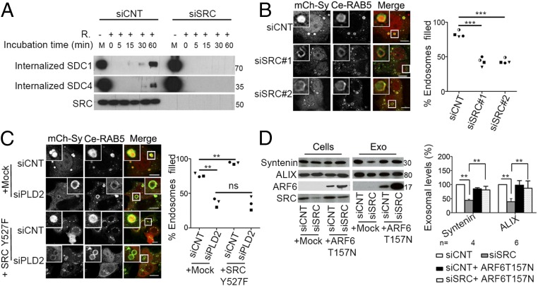
The cytoplasmic tyrosine kinase SRC controls cell growth, proliferation, adhesion, and motility. The current view is that SRC acts primarily downstream of cell-surface receptors to control intracellular signaling cascades. Here we reveal that SRC functions in cell-to-cell communication by controlling the biogenesis and the activity of exosomes. Exosomes are viral-like particles from endosomal origin that can reprogram recipient cells. By gain- and loss-of-function studies, we establish that SRC stimulates the secretion of exosomes having promigratory activity on endothelial cells and that syntenin is mandatory for SRC exosomal function. Mechanistically, SRC impacts on syndecan endocytosis and on syntenin-syndecan endosomal budding, upstream of ARF6 small GTPase and its effector phospholipase D2, directly phosphorylating the conserved juxtamembrane DEGSY motif of the syndecan cytosolic domain and syntenin tyrosine 46. Our study uncovers a function of SRC in cell-cell communication, supported by syntenin exosomes, which is likely to contribute to tumor-host interactions.

Keywords: ARF6; SRC; exosome; syndecan; syntenin.


Conflict of interest statement

The authors declare no conflict of interest.

Figures

Fig. 1.
Fig. 1.
SRC levels in MCF-7 donor cells determine the impact of exosomes on the migration of recipient HUVEC cells. (A) Conditioned media (CM) were collected from MCF-7 cells treated with nontargeting control RNAi (siCNT) or SRC RNAi (siSRC), grown in equal numbers for equal lengths of time. CM and corresponding CM that were depleted of exosomes by ultracentrifugation at 100,000 × g (CM-exo) were used to stimulate wound closures in monolayers of HUVEC cells. Closures are expressed as percentages, relative to the closure measured in the presence of DMEM (Control, taken as 100%). (B) Equivalent amounts of exosomes (50 µg as measured by Bradford assay) isolated from CM were used to stimulate wound closures. Exosomes were collected by ultracentrifugation and were resuspended in PBS. Cells were either depleted of SRC (siSRC) or treated with nontargeting RNAi (siCNT). Closures are expressed as percentages, relative to the closure measured in the presence of PBS (Control, taken as 100%) (ns, nonsignificant; *P < 0.05; **P < 0.01).
Fig. 2.
Fig. 2.
SRC regulates exosome number and cargo, and syntenin is mandatory for the promigratory activity of SRC-dependent exosomes. (AC) SRC effects on exosomes. Exosomes, isolated by ultracentrifugation, derived from control (siCNT) and SRC-depleted (siSRC) cells were analyzed by Western blot (A and C) using antibodies for several different markers, as indicated, or by NTA (B). Histograms represent signal intensities, mean ± SD, relative to signals measured in control samples (white bars), taken as 100% (**P < 0.01, ***P < 0.001). The dot plot represents the total number of particles, relative to the number measured in control samples, taken as 100%, with each independent experiment being represented by a different symbol (***P < 0.005). (D) SRC exosome promigratory effects require syntenin. Equivalent amounts of exosomes (40 µg measured by Bradford assay) isolated (by ultracentrifugation) from media conditioned by MCF-7 cells were used to stimulate the migration of HUVEC cells across the membrane of a Boyden chamber. MCF-7 cells were either “wild-type” or “syntenin-null” (clones 2b and 8b, obtained by gene inactivation, using CRISPR/Cas9) and were treated with various RNAi or transfected to overexpress SRC, as indicated. Migration is expressed as a percentage, relative to the number of cells migrating when the lower chamber is filled with DMEM/F12 medium not supplemented with exosomes but with PBS (taken as 100%), and is also compared with the migration measured when the lower chamber is filled with media containing 10% of FCS (not depleted of exosomes) (*P < 0.05).
Fig. 3.
Fig. 3.
SRC controls SDC endocytosis and syntenin endosomal budding upstream of ARF6–PLD2. (A) SDC internalization was monitored in reversible (by reduction, R) cell-surface biotinylation experiments. Western blots illustrate full-length SDC1 and SDC4 internalized over time and SRC depletion. M represents the membrane pool before internalization. Blots are representative of four independent SDC1 and two independent SDC4 experiments. (B) Confocal micrographs (Left) showing the accumulation of mCh-syntenin (mCh-Sy) inside the lumen of enlarged endosomes outlined by Ce-RAB5(Q79L) (Ce-RAB5) in control and SRC-depleted cells. (Scale bar, 10 µm.) Corresponding dot plot (Right) indicates the percentage of Ce-RAB5(Q79L) endosomes that is filled with mCh-syntenin, in the different conditions. Each quantification was performed considering at least 40–50 RAB5(Q79L) endosomes, in each experiment. (C) Confocal micrographs (Left) of MCF-7 cells that were treated with siCNT or PLD2 RNAi for 48 h and then transfected with expression vectors encoding SRC Y527F, Cerulean-RAB5(Q79L), and mCh-syntenin for 24 h. (Scale bar, 10 µm.) The dot plot (Right) represents the percentage of RAB5(Q79L) endosomes that is filled with mCh-syntenin, in the different conditions. (D) MCF-7 cells were treated with either SRC siRNA (siSRC) or nontargeting siRNA (siCNT) for 48 h and then transfected with an expression vector encoding the fast cycling mutant of ARF6 (ARF6-T157N) or mock-transfected with an empty vector. The cells and the exosomes produced by these cells were analyzed by Western blotting, using antibodies for several different markers, as indicated. Corresponding histograms (Right) represent signal intensities, mean ± SD, measured in exosomes, relative to signals measured in exosomes derived from the control cells (siCNT, followed by mock-transfection) (taken as 100%, white bars). All data were compiled from at least three independent experiments (ns, nonsignificant; **P < 0.01; ***P < 0.005).
Fig. 4.
Fig. 4.
SRC kinase activity and the tyrosine phosphorylations of both SDC and syntenin mediate SRC controls on endosomal budding and exosome release. (A) Equal numbers of MCF-7 cells, grown in the presence of serum, were left untreated (control) or treated with SRC inhibitor-1 (SI-1) or vehicle (DMSO). Proteins in exosomes (prepared by ultracentrifugation) were analyzed by Western blotting and quantified by densitometry, taking signals measured in exosomes derived from control MCF-7 cells as 100%. (B) Dot plot illustrating the total numbers of particles present in exosomal preparations from cells treated with SRC inhibitor-1, relative to the values obtained for preparations originating from cells treated with DMSO (taken as 100%), as determined by NTA. (C and D) Dot plot indicating the percentage of Ce-RAB5(Q79L) endosomes filled with SDC1 ICD, in MCF-7 cells transfected with empty expression vector (Mock), and in cells overexpressing WT or various tyrosine mutant forms of SDC1. Note that cells are either grown in serum (C) or starved for 24 h (D), before fixation. (E) Dot plot indicating the percentage of Ce-RAB5(Q79L) endosomes filled with mCh-syntenin, in MCF-7 cells overexpressing WT or various tyrosine mutant forms of mCh-syntenin, and starved for 24 h before fixation. (F) Dot plot indicating the percentage of Ce-RAB5(Q79L) endosomes filled with mCh-syntenin, in MCF-7 cells expressing either wild-type or phosphomimetic forms of SDC1 and mCh-syntenin, and treated with SRC inhibitor or vehicle (DMSO) in the combinations indicated. Cells were starved for 24 h before fixation. Each quantification (DF) was performed considering at least 40–50 RAB5(Q79L) endosomes, in each experiment (ns, nonsignificant; *P < 0.05; **P < 0.01; ***P < 0.005).
Fig. 5.
Fig. 5.
Model recapitulating the relation between SRC kinase and the SDC–syntenin exosomal pathway, as revealed in the present study. (A) The conditioned medium of MCF-7 cells has promigratory effects on HUVEC cells. This activity is mediated by exosomes and is lost upon SRC depletion and increased upon SRC gain of function in exosome donor cells. In MCF-7 cells, syntenin is mandatory for SRC promigratory effects on HUVEC cells. (B) Processes under the control of SRC. SRC acts by favoring SDC internalization and SDC–syntenin endosomal budding (upstream of ARF6/PLD2). SRC is directly phosphorylating the conserved tyrosine in the membrane proximal DEGSY motif of SDC and thereby increases the recruitment and binding of syntenin at the endosomal membrane. However, the phosphorylation of syntenin by SRC, on the tyrosine at position 46, contributes to endosomal budding and exosome release. SRC thereby influences the number of exosomes that is released and controls the loading of exosomes with cargo such as integrins and receptor tyrosine kinases, known partners of SDCs.

References

    1. Sirvent A, Benistant C, Roche S. Oncogenic signaling by tyrosine kinases of the SRC family in advanced colorectal cancer. Am J Cancer Res. 2012;2:357–371. - PMC - PubMed
    1. Wheeler DL, Iida M, Dunn EF. The role of Src in solid tumors. Oncologist. 2009;14:667–678. - PMC - PubMed
    1. Yeatman TJ. A renaissance for SRC. Nat Rev Cancer. 2004;4:470–480. - PubMed
    1. Okada M. Regulation of the SRC family kinases by Csk. Int J Biol Sci. 2012;8:1385–1397. - PMC - PubMed
    1. Mineo M, et al. Exosomes released by K562 chronic myeloid leukemia cells promote angiogenesis in a Src-dependent fashion. Angiogenesis. 2012;15:33–45. - PMC - PubMed

Publication types

MeSH terms