. 2017 Nov 21;114(47):12448-12453.
doi: 10.1073/pnas.1707925114. Epub 2017 Nov 6.

Blockade of activin type II receptors with a dual anti-ActRIIA/IIB antibody is critical to promote maximal skeletal muscle hypertrophy

Affiliations

Blockade of activin type II receptors with a dual anti-ActRIIA/IIB antibody is critical to promote maximal skeletal muscle hypertrophy

Frederic Morvan et al. Proc Natl Acad Sci U S A. .

Abstract

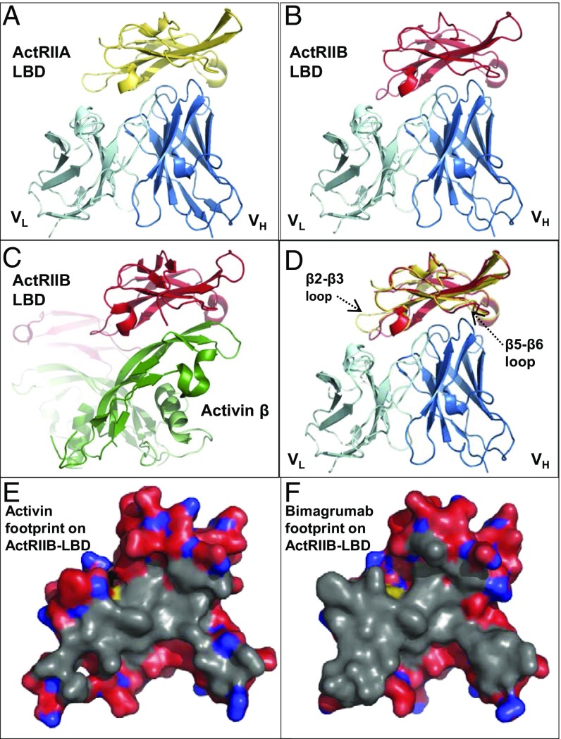
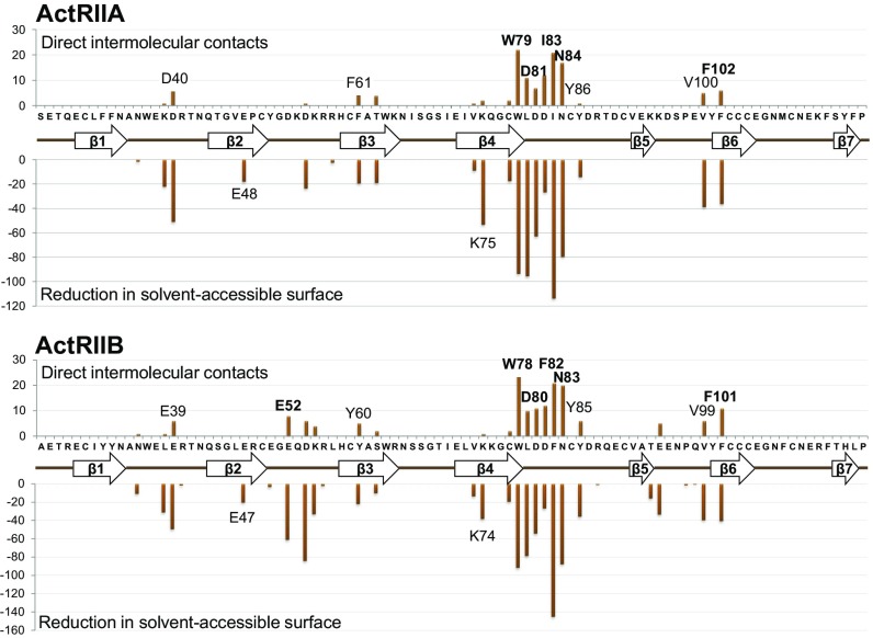
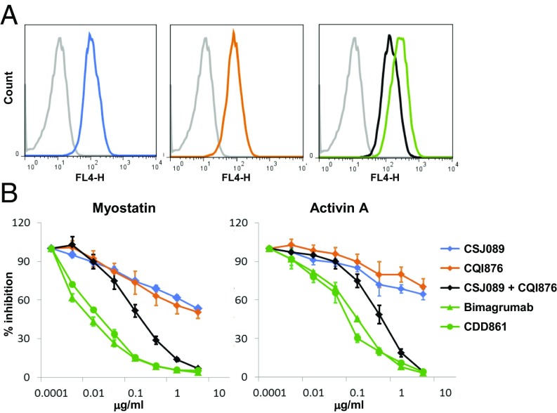
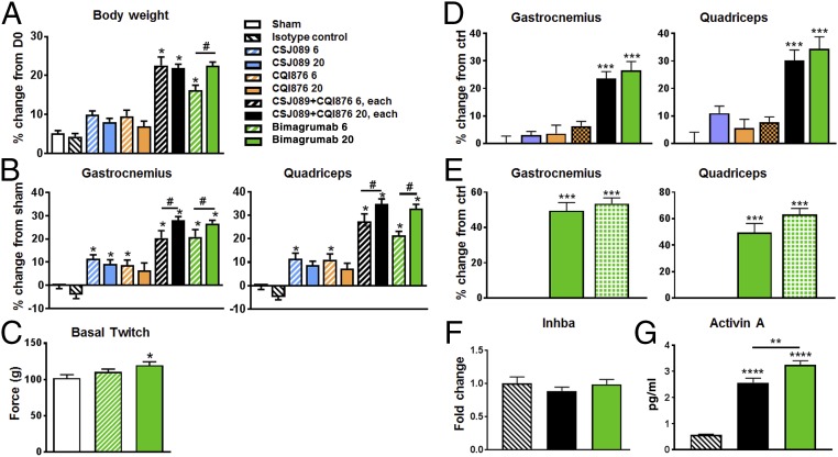
The TGF-β family ligands myostatin, GDF11, and activins are negative regulators of skeletal muscle mass, which have been reported to primarily signal via the ActRIIB receptor on skeletal muscle and thereby induce muscle wasting described as cachexia. Use of a soluble ActRIIB-Fc "trap," to block myostatin pathway signaling in normal or cachectic mice leads to hypertrophy or prevention of muscle loss, perhaps suggesting that the ActRIIB receptor is primarily responsible for muscle growth regulation. Genetic evidence demonstrates however that both ActRIIB- and ActRIIA-deficient mice display a hypertrophic phenotype. Here, we describe the mode of action of bimagrumab (BYM338), as a human dual-specific anti-ActRIIA/ActRIIB antibody, at the molecular and cellular levels. As shown by X-ray analysis, bimagrumab binds to both ActRIIA and ActRIIB ligand binding domains in a competitive manner at the critical myostatin/activin binding site, hence preventing signal transduction through either ActRII. Myostatin and the activins are capable of binding to both ActRIIA and ActRIIB, with different affinities. However, blockade of either single receptor through the use of specific anti-ActRIIA or anti-ActRIIB antibodies achieves only a partial signaling blockade upon myostatin or activin A stimulation, and this leads to only a small increase in muscle mass. Complete neutralization and maximal anabolic response are achieved only by simultaneous blockade of both receptors. These findings demonstrate the importance of ActRIIA in addition to ActRIIB in mediating myostatin and activin signaling and highlight the need for blocking both receptors to achieve a strong functional benefit.

Keywords: ActRII; activin; dual antibody; hypertrophy; myostatin.


Conflict of interest statement

Conflict of interest statement: All authors but F.W. are employees of Novartis Pharma AG (F.M., C.Z., C.S., J.-M.R., C.J., A.S., P.B., V.R., G.T., M.K., X.L., S.L., E.K., G.M., C.K., S.G., D.J.G., E.L.-T.), and some are also shareholders of Novartis. F.W. was an employee of MorphoSys AG at the time of contribution.

Figures

Fig. 1.
Fig. 1.
Binding modalities of bimagrumab to ActRIIA and ActRIIB. Crystal structures of the bimagrumab Fv (light/dark blue ribbon) complex with (A) human ActRIIA LBD (gold ribbon) or (B) human ActRIIB LBD (red ribbon), shown in the same orientation. (C) Crystal structure of the mouse ActRIIB LBD complex with human activin-β (PDB entry 1S4Y) (35), shown in the same orientation as in A, B, and D. (D) Overlay of the ActRIIA Fv complex (gold ribbon) and ActRIIB Fv complex (red ribbon). (E) Footprint of activin (gray surface, calculated from PDB entry 1S4Y) and (F) bimagrumab on the ActRIIB LBD. Note the extensive overlap between the two binding surfaces.
Fig. 2.
Fig. 2.
ActRIIA (Upper) and ActRIIB (Lower) epitope recognized by bimagrumab. The Upper part of each panel shows the number of direct intermolecular contacts between nonhydrogen atoms within a 4.0-Å distance; Lower part shows the reduction in solvent-accessible surface upon complex formation. The amino acid sequence of the respective ActRII LBD is displayed on the horizontal axis, together with a schematic representation of the secondary structure elements (arrows, β-strands; thick lines, connecting loops).
Fig. 3.
Fig. 3.
Cellular responses to ActRIIA and ActRIIB neutralization. (A) Binding of anti-ActRIIA (CSJ089, blue), anti-ActRIIB (CQI876, orange), anti-ActRIIA and ActRIIB Abs combination (black), bimagrumab (green), or isotype control Ab (gray) to HEK293T/17 cells. (B) Efficacy/potency of antibodies or combination thereof at blocking myostatin or activin A-induced Smad2/3 response. Single-specificity Abs (CSJ089, blue), (CQI876, orange), or combination thereof (CSJ089 and CQI876, black), or bimagrumab (green, triangle) or CDD861 (green, circle) were tested for their ability to inhibit myostatin (10 ng/mL) or activin A (10 ng/mL)-induced Smad2/3 response in a CAGA-luciferase reporter gene assay in HEK293T/17 cells.
Fig. 4.
Fig. 4.
Hypertrophy response as measured via (A) body weight, (B) muscle weight change from sham group, after 4-wk treatment with ActRIIA-specific Ab, ActRIIB-specific Ab, combination thereof, and bimagrumab in SCID mice (n = 12 per group). Mice were untreated, sham group (white), or treated with weekly s.c. injection of isotype control antibody (20 mg/kg/wk) or of anti-ActRIIA Ab (CSJ089, blue, 6 or 20 mg/kg), an anti-ActRIIB Ab (CQI876, orange, 6 or 20 mg/kg), a combination of CSJ089 and CQI876 (black, 6 or 20 mg/kg of each Ab), or bimagrumab (green, 6 or 20 mg/kg). (C) Invasive muscle contractile function determination in gastrocnemius muscle of sham (white) and bimagrumab (green, 6 and 20 mg/kg)-treated groups, average of three stimulations. Hypertrophy response was measured through gastrocnemius and quadriceps muscle weight changes in SCID mice, (D) after 2-wk treatment with the same antibodies as in A, all dosed at 20 mg/kg, with CQI876 being also dosed at 100 mg/kg (orange crosses), (E) after 4-wk treatment with weekly s.c. injection of isotype control antibody (stripes), dual anti-ActRIIA/ActRIIB Ab (CDD861, green pattern, 20 mg/kg), and bimagrumab (green, 20 mg/kg). (F) Activin A (inhba gene) expression changes in gastrocnemius muscle of SCID mice, after 2-wk treatment with isotype control, combination of anti-ActRIIA and anti-ActRIIB Abs (black), or bimagrumab (green) as in D. (G) ELISA data for activin A level in serum from mice of D/F. Data are presented as mean ± SEM analyzed using one-way ANOVA; differences vs. control were considered statistically significant, *P < 0.05; **P < 0.01; ***P < 0.001; ****P < 0.0001. Unpaired t test 20 vs. 6 mg/kg, #P < 0.05.

Comment in

References

    1. Egerman MA, Glass DJ. Signaling pathways controlling skeletal muscle mass. Crit Rev Biochem Mol Biol. 2014;49:59–68. - PMC - PubMed
    1. Bodine SC. Disuse-induced muscle wasting. Int J Biochem Cell Biol. 2013;45:2200–2208. - PMC - PubMed
    1. Glass D, Roubenoff R. Recent advances in the biology and therapy of muscle wasting. Ann N Y Acad Sci. 2010;1211:25–36. - PubMed
    1. Morley JE. Sarcopenia in the elderly. Fam Pract. 2012;29:i44–i48. - PubMed
    1. Han HQ, Zhou X, Mitch WE, Goldberg AL. Myostatin/activin pathway antagonism: Molecular basis and therapeutic potential. Int J Biochem Cell Biol. 2013;45:2333–2347. - PubMed

MeSH terms