. 2017 Nov 6;7(1):14520.
doi: 10.1038/s41598-017-15163-5.

Neuroprotective effect of Ruminococcus albus on oxidatively stressed SH-SY5Y cells and animals

Affiliations

Neuroprotective effect of Ruminococcus albus on oxidatively stressed SH-SY5Y cells and animals

Jieun Park et al. Sci Rep. .

Abstract

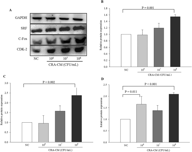
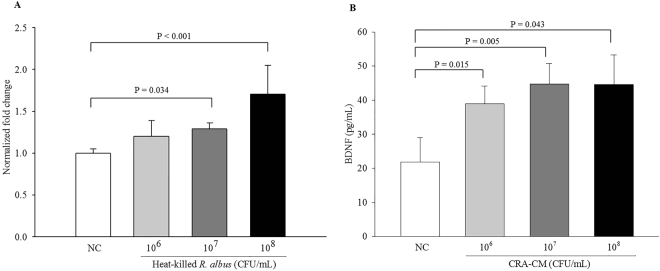
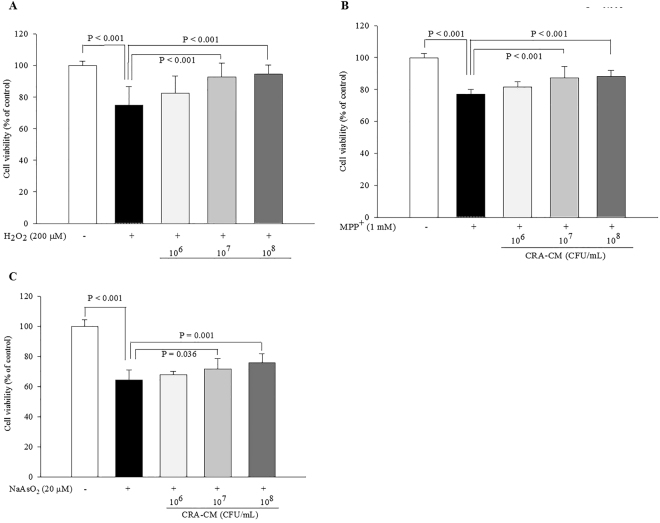
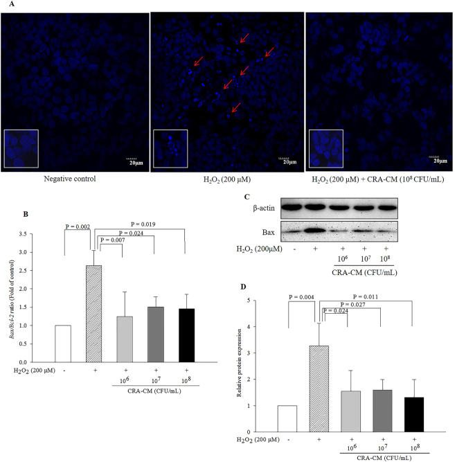
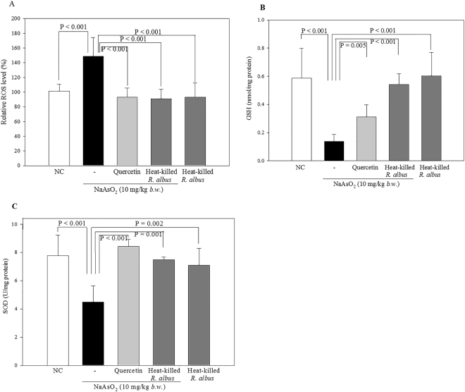
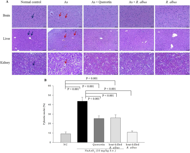
Recent evidence shows that the gut microbiota has an important role in gut-brain crosstalk and is linked to neuronal disorders. The aim of this study was to investigate the effects of intestinal Ruminococcus albus with probiotic potential on neuroprotection in oxidatively stressed SH-SY5Y neuroblastoma cells and animals. To investigate these effects, conditioned medium was prepared using Caco-2 cells cultured with heat-killed R. albus (CRA-CM). Caco-2 cells cultured with heat-killed R. albus showed increased BDNF expression and BDNF protein levels increased in CRA-CM. CRA-CM up-regulated the protein expression levels of SRF, C-fos and CDK2. In addition, CRA-CM protected SH-SY5Y cells from H2O2-induced cell death. CRA-CM significantly decreased the Bax/Bcl-2 ratio in oxidatively stressed SH-SY5Y cells. Animal experiments showed that oral administration of heat-killed R. albus for 15 days attenuated the oxidative stress induced by sodium arsenate. Treatment with heat-killed R. albus reduced the level of ROS, and the levels of SOD and GSH increased in oxidatively stressed brains. In conclusion, the secretome prepared from Caco-2 cells cultured with heat-killed R. albus might promote neuronal proliferation through the activation of cell proliferation-related proteins, and heat-killed R. albus protects neurons from oxidative damage by reducing ROS levels and increasing SOD and GSH levels.


Conflict of interest statement

The authors declare that they have no competing interests.

Figures

Figure 1
Figure 1
Effects of CRA-CM on cell viability in SH-SY5Y cells. SH-SY5Y cells were treated with CRA-CM for 24 h. Cell viability was measured by MTT (A) and LDH (B) assay. The data are representative of three independent experiments performed in triplicate.
Figure 2
Figure 2
Effects of CRA-CM on the expression of SRF, C-fos and CDK2 in SH-SY5Y cells. The expression levels of SRF, C-fos and CDK2 were measured by western blotting and cropped blots are displayed (A), and the densities of SRF (B), c-Fos (C) and CDK2 (D) were quantified. The full-length blots are shown in Supplementary Fig. S2. The data are expressed as the mean ± SD of three independent experiments performed in triplicate.
Figure 3
Figure 3
mRNA expression of BDNF in Caco-2 cells treated with heat-killed R. albus (A) and BDNF levels in CRA-CM (B). The relative levels of mRNA expression were normalized using GAPDH as an internal control. BDNF protein levels in CRA-CM were measured by ELISA. The negative control was treated with PBS. The data are representative of three independent experiments performed in triplicate.
Figure 4
Figure 4
Effects of CRA-CM on expression of β-tubulin in SH-SY5Y cells. Expression levels of β-tubulin were measured by western blotting and cropped blots are displayed (A), and quantified (B). The full-length blots are shown in Supplementary Fig. S3. The data are expressed as the mean ± SD of three independent experiments performed in triplicate.
Figure 5
Figure 5
Neuroprotective effects of CRA-CM on cell viability in oxidatively stressed SH-SY5Y cells. Oxidative stress was induced by (A) H2O2 (200 μM), (B) MPP+ (1 mM) or (C) NaAsO2 (20 μM). SH-SY5Y cells were pretreated with CRA-CM. After 4 h, H2O2, MPP+ or NaAsO2 was applied for 20 h. Cell viability was measured using an MTT assay. The data are representative of three independent experiments performed in triplicate.
Figure 6
Figure 6
Protective effects of CRA-CM against fragmentation of nuclei in oxidatively stressed SH-SY5Y cells. (A) Hoechst 33258 (5 μg/mL) staining was performed in SH-SY5Y cells. Apoptotic cells were identified by the condensation and fragmentation of their nuclei (red arrow). The negative control Caco-2 cells were treated with PBS. The expression levels of apoptosis-related genes and proteins (Bcl-2 and Bax) in oxidatively stressed SH-SY5Y cells were measured by qPCR (B) and western blotting and cropped blots are displayed (C), respectively. Western blot analysis was quantified using β-actin as an internal control (D). The full-length blots are shown in Supplementary Fig. S4. The data are representative of three independent experiments performed in triplicate.
Figure 7
Figure 7
Effect of heat-killed R. albus on ROS levels and antioxidant activity in brain homogenates of oxidatively stressed rats. Sodium arsenate (10 mg/kg) was used to induce oxidative stress in the rat brain. The ROS levels were evaluated using the dichlorofluorescein diacetate (DCF-DA) method (A). GSH levels of brain tissues in rats (B). SOD activity levels of brain tissue in rats (C). The data are expressed as the mean ± SD.
Figure 8
Figure 8
Histological analysis of brain, liver, and kidneys of rats oxidatively stressed with sodium arsenate (As). (A) All images were captured at 100× magnification except the brain images (200×). The blue and red arrows show normal and damaged tissues, respectively. (B) The percent of pyknotic nuclei per field (140 μm2) in brain (n = 5).

References

    1. Dauer W, Przedborski S. Parkinson’s disease: mechanisms and models. Neuron. 2003;39:889–909. doi: 10.1016/S0896-6273(03)00568-3. - DOI - PubMed
    1. Schapira AH, Jenner P. Etiology and pathogenesis of Parkinson’s disease. Movement Disorders. 2011;26:1049–1055. doi: 10.1002/mds.23732. - DOI - PubMed
    1. Jenner P. Oxidative stress in Parkinson’s disease. Annals of Neurology. 2003;53(Suppl 3):S26–S36. doi: 10.1002/ana.10483. - DOI - PubMed
    1. Jenner P, Olanow CW. The pathogenesis of cell death in Parkinson’s disease. Neurology. 2006;66:S24–S36. doi: 10.1212/WNL.66.10_suppl_4.S24. - DOI - PubMed
    1. Ye M, Kim M, Bae H. Neuro-protective effects of Ligustri Fructus by suppression of oxidative stress in mouse model of Parkinson’s disease. Oriental Pharmacy and Experimental Medicine. 2016;16:123–129. doi: 10.1007/s13596-016-0223-0. - DOI

Publication types

MeSH terms