. 2017 Aug 23;7(15):3803-3813.
doi: 10.7150/thno.21068. eCollection 2017.

A H2O2-Responsive Theranostic Probe for Endothelial Injury Imaging and Protection

Affiliations

A H2O2-Responsive Theranostic Probe for Endothelial Injury Imaging and Protection

Cheng-Kun Wang et al. Theranostics. .

Abstract

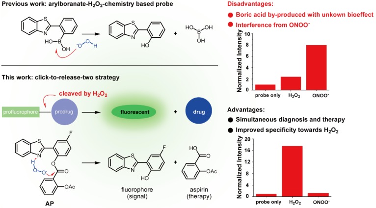
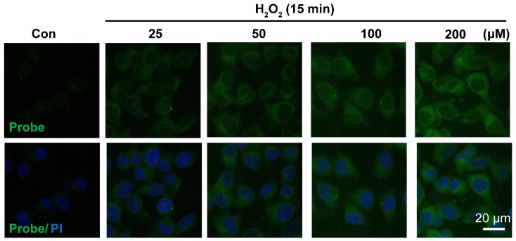
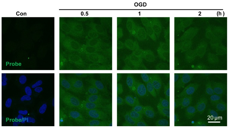
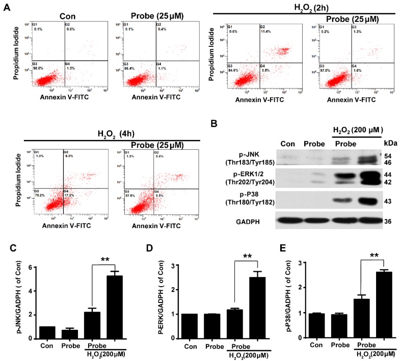
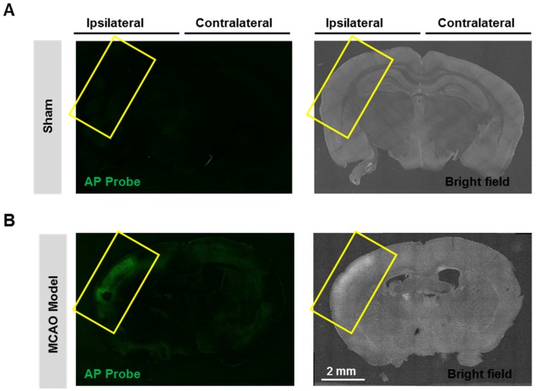
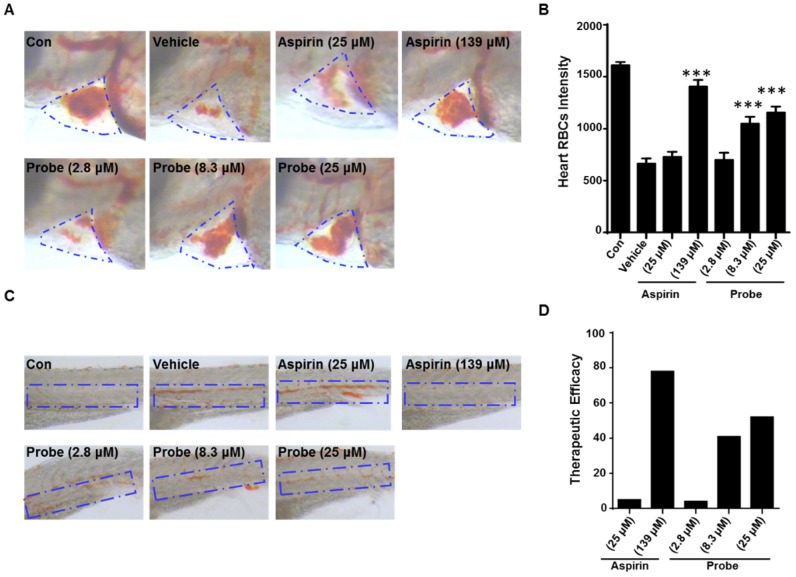
Overproduction of H2O2 causes oxidative stress and is the hallmark of vascular diseases. Tracking native H2O2 in the endothelium is therefore indispensable to gain fundamental insights into this pathogenesis. Previous fluorescent probes for H2O2 imaging were generally arylboronates which were decomposed to emissive arylphenols in response to H2O2. Except the issue of specificity challenged by peroxynitrite, boric acid by-produced in this process is actually a waste with unknown biological effects. Therefore, improvements could be envisioned if a therapeutic agent is by-produced instead. Herein, we came up with a "click-to-release-two" strategy and demonstrate that dual functional probes could be devised by linking a fluorophore with a therapeutic agent via a H2O2-responsive bond. As a proof of concept, probe AP consisting of a 2-(2'-hydroxyphenyl) benzothiazole fluorophore and an aspirin moiety has been prepared and confirmed for its theranostic effects. This probe features high specificity towards H2O2 than other reactive species including peroxynitrite. Its capability to image and ameliorate endothelial injury has been verified both in vitro and in vivo. Noteworthy, as a result of its endothelial-protective effect, AP also works well to reduce thrombosis formation in zebrafish model.

Keywords: endothelial injury; fluorescent imaging; hydrogen peroxide; oxidative stress.; theranostic probe.


Conflict of interest statement

Competing Interests: The authors have declared that no competing interest exists.

Figures

Figure 1
Figure 1
Comparison of previous arylboranate-H2O2 chemistry-based probe, and probe AP in this work. For the same fluorophore, boric acid-based probe showed poor selectivity for H2O2, while aspirin-based probe AP was almost immune to ONOO-. Data were the normalized fluorescent intensity of the probes (10 μM) at 460 nm before or after the treatment of H2O2 (500 μM) or ONOO- (20 μM) for 30 min.
Figure 2
Figure 2
Photophysical responses of AP to H2O2. A) UV-Vis absorption response of AP (30 μM) towards H2O2 (1.0 mM) as time lapsed. B) Fluorescence response of AP (10 μM) towards H2O2 (200 μM) as time lapsed. C) Fluorescence response of AP (10 μM) towards various concentrations of H2O2 at a time point of 30 min. D) Fluorescent responses of AP (10 μM) toward various analytes (200 μM) after a reaction time of 30 min, where F represents the fluorescent intensity of AP at 476 nm after the treatment of various analytes, and F0 represents the intensity of blank AP solution at 476 nm. (1) probe blank, (2) H2O2, (3) 1O2, (4) O2-, (5) ClO-, (6) THBP, (7) ONOO-, (8) NO, (9) NO2-, (10) NO3-, (11) GSH, (12) Cys, (13) Hcy, (14) Gly, (15) Ala, (16) Mg2+, (17) Ca2+, (18) Fe3+, (19) Fe2+, (20) K+, (21) Cu2+, (22) Zn2+. All fluorescence data were collected in PBS (pH 7.4, 100 mM) at 37oC with λex 375 nm. UV data were obtained in PBS with 50% EtOH as co-solvent.
Figure 3
Figure 3
Representative confocal images showed dose-dependent increase of AP fluorescence in endothelial cells. Cells were seeded on 24-well glass cover slips overnight and then pre-incubated with AP (5.0 μM) for 15 min, followed by challenging without or with H2O2 (25-200 μM) for 15 min. AP fluorescence (green, 460 nm) was excited at 405 nm. PI counterstaining indicated nuclear localization (blue, 620 nm). All images were captured using a Nikon A1R confocal microscope with the same settings. Overlay image of all captured fluorescence intensities are shown. Scale bar represents 20 μm.
Figure 4
Figure 4
Visualizing endogenous H2O2 formation using AP in endothelial cells following oxygen-glucose deprivation (OGD). The representative confocal images showed temporal changes of AP fluorescence (green, 460 nm) in EA.hy926 endothelial cells over 0.5 to 2 h following OGD. PI counterstaining indicated nuclear localization (blue, 620 nm). Overlay image of AP fluorescence and PI are shown. Scale bar represents 20 μm.
Figure 5
Figure 5
The protective role of AP against H2O2-induced endothelial apoptosis. A) The apoptosis of endothelial cells was labelled with annexin V-FITC/propidium iodide (PI) and determined using flow cytometry. EA.hy926 cells were seeded on 12-well plates overnight and then pre-incubated with AP (25 μM) for 15 min, followed by stimulation with H2O2 (200 μM, 2 h or 4 h) in HBSS medium. B) The representative blots for phosphorylation of JNK, ERK and p38 in the presence of AP probe upon H2O2 (200 μM, 12 h) exposure in DMEM medium. Summary of phospho-JNK (C), phospho-ERK (D) and phospho-p38 (E) were indicated as densitometric values. **p < 0.01.
Figure 6
Figure 6
Visualizing endogenous H2O2 formation using AP in MCAO mouse. The representative confocal images showed changes of AP fluorescence (green) in brain ischemic region in MCAO mice. Insert frame: Brain ischemic region. Scale bar represents 2 mm.
Figure 7
Figure 7
The antithrombotic effect of probe AP. Zebrafish treated with arachidonic acid for 1.5 h were used as thrombosis model. A) Effects of probe AP on heart RBCs in thrombotic zebrafish. The heart red blood cells were stained with o-dianisidine staining. The heart RBCs intensity of zebrafish increased following AP probe treatment (1.5 h) in the presence of arachidonic acid (80 µM). B) Quantification data for Figure A. C) Effect of AP probe on thrombus formation in the caudal vein under a dissecting stereomicroscope. Images from aspirin or AP probe treatment are shown from a representative caudal vein of zebrafish from each group. Thrombus formation was markedly reduced in zebrafish treated with AP probe (1.5 h) in the presence of arachidonic acid. D) The therapeutic efficacy of AP probe on thrombosis formation.

References

    1. Vanhoutte PM, Shimokawa H, Tang EH, Feletou M. Endothelial dysfunction and vascular disease. Acta Physiol (Oxf) 2009;196:193–222. - PubMed
    1. Kasote DM, Hegde MV, Katyare SS. Mitochondrial dysfunction in psychiatric and neurological diseases: cause(s), consequence(s), and implications of antioxidant therapy. Biofactors. 2013;39:392–406. - PubMed
    1. McCann SK, Roulston CL. NADPH Oxidase as a Therapeutic Target for Neuroprotection against Ischaemic Stroke: Future Perspectives. Brain Sci. 2013;3:561–98. - PMC - PubMed
    1. Schriewer JM, Peek CB, Bass J, Schumacker PT. ROS-mediated PARP activity undermines mitochondrial function after permeability transition pore opening during myocardial ischemia-reperfusion. J Am Heart Assoc. 2013;2:e159. - PMC - PubMed
    1. Hao XL, Kang Y, Li JK, Li QS, Liu EL, Liu XX. Protective effects of hyperoside against H2O2-induced apoptosis in human umbilical vein endothelial cells. Mol Med Rep. 2016;14:399–405. - PubMed

Publication types

LinkOut - more resources