

Case Reports
doi: 10.1155/2017/2595036.
Epub 2017 Oct 3.
Unusual Etiology and Diagnosis of Oroantral Communication due to Late Implant Failure
Affiliations
- PMID: 29109871
- PMCID: PMC5646311
- DOI: 10.1155/2017/2595036
Item in Clipboard
Case Reports
Unusual Etiology and Diagnosis of Oroantral Communication due to Late Implant Failure
Case Rep Dent.
2017.
Abstract
Oroantral communication (OAC) rarely occurs long after implant placement. The present report describes the rare etiology and the difficulty of the diagnosis of an uncommon OAC occurring 10 years after the implant placement in the posterior maxilla. The difficulty of the diagnosis lies in the absence of clinical symptoms of sinusitis and presence of multiunit prosthesis hiding implant failure. This case report supports the need for sinus check-up during a routine implant examination.
Figures
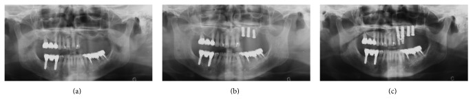
Placement of implants, panoramic radiographs. (a) Initial situation, (b) immediately after implant placement in sites 23, 24, and 25, and (c) 7 months later, immediately after implant placement in sites 25 and 26.
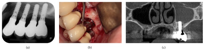
Ten years after implant placement. (a) Periapical radiograph, (b) clinical view, flap at site 25, and (c) cone-beam computed tomography image. Note the opacity of the left sinus.
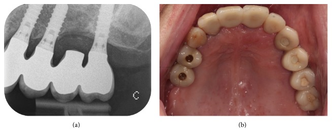
The implant 25 was removed and the bridge was rescrewed. The patient underwent antibiotic treatment. (a) Periapical radiograph and (b) clinical view.
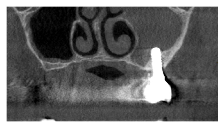
Six months after the removal of the implant 25. The cone-beam computed tomography image revealed that the opacity of the left sinus was still present.
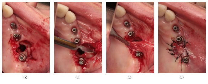
The oroantral fistula was closed with a buccal advancement flap. (a)–(d) Clinical views.
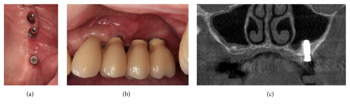
Two months after the closure of OAF. (a)-(b) Clinical views. The oroantral fistula remained successfully closed. (c) Cone-beam computed tomography image. The left sinus was totally healed.
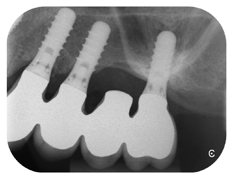
Two years after the closure of OAF; the radiographic control showed a stable crestal bone level.
Similar articles
-
Concomitant oroantral communication repair and immediate implant placement: a five-year case report.Implant Dent. 2008 Jun;17(2):176-81. doi: 10.1097/ID.0b013e318166dbe7. Implant Dent. 2008. PMID: 18545049
-
Simultaneous oroantral communication closure, sinus-lifting, and particulate bone grafting and immediate dental implant perforation.Niger J Clin Pract. 2016 Jul-Aug;19(4):556-558. doi: 10.4103/1119-3077.183300. Niger J Clin Pract. 2016. PMID: 27251977
-
Disappearance of a dental implant after migration into the maxillary sinus: an unusual case.J Korean Assoc Oral Maxillofac Surg. 2015 Oct;41(5):278-80. doi: 10.5125/jkaoms.2015.41.5.278. Epub 2015 Oct 20. J Korean Assoc Oral Maxillofac Surg. 2015. PMID: 26568932 Free PMC article.
-
Decision-making in closure of oroantral communication and fistula.Int J Implant Dent. 2019 Apr 1;5(1):13. doi: 10.1186/s40729-019-0165-7. Int J Implant Dent. 2019. PMID: 30931487 Free PMC article. Review.
-
Oroantral communication.Oral Maxillofac Surg Clin North Am. 2012 May;24(2):239-47, viii-ix. doi: 10.1016/j.coms.2012.01.015. Oral Maxillofac Surg Clin North Am. 2012. PMID: 22503070 Review.
Cited by
-
Oroantral communication, its causes, complications, treatments and radiographic features: A pictorial review.Imaging Sci Dent. 2021 Sep;51(3):307-311. doi: 10.5624/isd.20210035. Epub 2021 Jul 13. Imaging Sci Dent. 2021. PMID: 34621658 Free PMC article. Review.
-
Management of Oro-Antral Communication: A Systemic Review of Diagnostic and Therapeutic Strategies.Diagnostics (Basel). 2025 Jan 16;15(2):194. doi: 10.3390/diagnostics15020194. Diagnostics (Basel). 2025. PMID: 39857078 Free PMC article. Review.
References
-
- Franco-Carro B., Barona-Dorado C., Martínez-González M. J. S., Rubio-Alonso L. J., Martínez-González J. M. Meta-analytic study on the frequency and treatment of oral antral communications. Medicina Oral, Patologia Oral y Cirugia Bucal. 2011;16(5):e682–e687. doi: 10.4317/medoral.17058. - DOI - PubMed
Publication types
LinkOut - more resources
Full Text Sources
Other Literature Sources
