

Are All Retinal Nerve Fiber Layer Defects on Optic Coherence Tomography Glaucomatous?
- PMID: 29109895
- PMCID: PMC5661176
- DOI: 10.4274/tjo.86461
Are All Retinal Nerve Fiber Layer Defects on Optic Coherence Tomography Glaucomatous?
Abstract
Objectives: In this study, we investigated the patients who were referred to our clinic with a prediagnosis of glaucoma based on retinal nerve fiber layer (RNFL) defects on optic coherence tomography (OCT) but were determined to have nonglaucomatous RNLF defects upon detailed examination.
Materials and methods: The ophthalmic examination notes, OCT images, Heidelberg retinal tomography (HRT) II and fundus photographs of 357 patients were retrospectively evaluated. Final diagnoses of these patients were investigated.
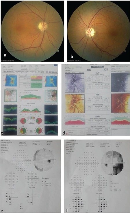
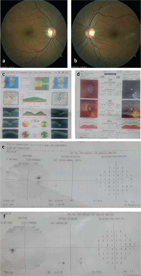
Results: Of the 357 patients, 216 (60.5%) were diagnosed as open angle glaucoma, 33 (9.2%) as low-tension glaucoma, 39 (10.9%) as pre-perimetric glaucoma. The ophthalmic examinations of 14 patients (3.9%) were normal and there were no RNFL defects in OCT examinations after dilatation. In 39 patients (10.9%), the ophthalmic and optic disc examinations were completely normal and no etiologic factor explaining RNFL defects was found. Twenty-two eyes of 16 patients (4.5%) were included in this study (the mean age was 53.8±11.5 years; 9 men and 7 women). After detailed questioning of the medical history and systemic and neurologic examinations, a diagnosis of ischemic optic neuropathy was made in 11 eyes (10 patients) (2.8%), optic neuritis in 3 eyes (2 patients) (0.6%), optic disc drusen in 4 eyes (2 patients) (0.6%), pseudotumor cerebri in 2 eyes (1 patient) (0.3%), and cerebral palsy in 2 eyes (1 patient) (0.3%).
Conclusion: Decrease in RNFL thickness on OCT images alone may be misleading in glaucoma examination. In cases where optic disc cupping is not evident, diagnosis should not be based on OCT RNFL examinations alone, and the patient's medical history, detailed ophthalmic examination, OCT optic disc parameters, HRT, and visual field tests should all be carefully evaluated together.
Keywords: Anterior ischemic optic neuropathy; Retinal nerve fiber layer; glaucoma; optic coherence tomography.
Conflict of interest statement
Conflict of Interest: No conflict of interest was declared by the authors.
Figures
References
-
- Schuman JS, Hee MR, Arya AV, Pedut-Kloizman T, Puliafito CA, Fujimoto JG, Swanson EA. Optical coherence tomography: a new tool for glaucoma diagnosis. Curr Opin Ophthalmol. 1995;6:89–95. - PubMed
-
- Puliafito CA, Hee MR, Lin CP, Reichel E, Schuman JS, Duker JS, Izatt JA, Swanson EA, Fujimoto JG. Imaging of macular diseases with optical coherence tomography. Ophthalmology. 1995;102:217–229. - PubMed
-
- Hee MR, Puliafito CA, Wong C, Duker JS, Reichel E, Schuman JS, Swanson EA, Fujimoto JG. Optical coherence tomography of macular holes. Ophthalmology. 1995;102:748–756. - PubMed
LinkOut - more resources
Full Text Sources
Other Literature Sources