

Limbal Stem Cell Deficiency and Treatment with Stem Cell Transplantation
- PMID: 29109898
- PMCID: PMC5661179
- DOI: 10.4274/tjo.72593
Limbal Stem Cell Deficiency and Treatment with Stem Cell Transplantation
Abstract
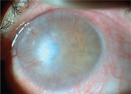
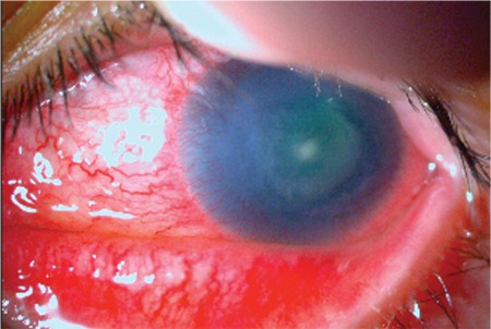
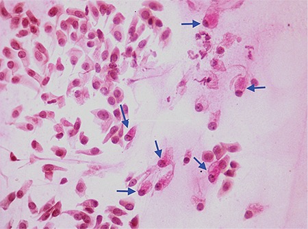
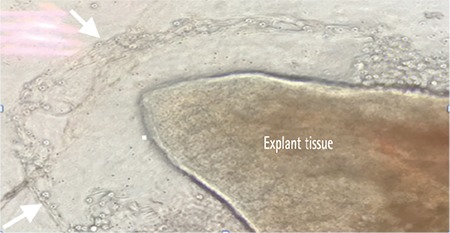
The cornea is the outermost tissue of the eye and it must be transparent for the maintenance of good visual function. The superficial epithelium of the cornea, which is renewed continuously by corneal stem cells, plays a critical role in the permanence of this transparency. These stem cells are localized at the cornea-conjunctival transition zone, referred to as the limbus. When this zone is affected/destroyed, limbal stem cell deficiency ensues. Loss of limbal stem cell function allows colonization of the corneal surface by conjunctival epithelium. Over 6 million people worldwide are affected by corneal blindness, and limbal stem cell deficiency is one of the main causes. Fortunately, it is becoming possible to recover vision by autologous transplantation of limbal cells obtained from the contralateral eye in unilateral cases. Due to the potential risks to the donor eye, only a small amount of tissue can be obtained, in which only 1-2% of the limbal epithelial cells are actually limbal stem cells. Vigorous attempts are being made to expand limbal stem cells in culture to preserve or even enrich the stem cell population. Ex vivo expanded limbal stem cell treatment in limbal stem cell deficiency was first reported in 1997. In the 20 years since, various protocols have been developed for the cultivation of limbal epithelial cells. It is still not clear which method promotes effective stem cell viability and this remains a subject of ongoing research. The most preferred technique for limbal cell culture is the explant culture model. In this approach, a small donor eye limbal biopsy is placed as an explant onto a biocompatible substrate (preferably human amniotic membrane) for expansion. The outgrowth (cultivated limbal epithelial cells) is then surgically transferred to the recipient eye. Due to changing regulations concerning cell-based therapy, the implementation of cultivated limbal epithelial transplantation in accordance with Good Laboratory Practice using xenobiotic-free systems is becoming widely accepted both in Turkey and worldwide.
Keywords: Limbal stem cell deficiency; cultured cells; stem cell transplantation.
Conflict of interest statement
Conflict of Interest: No conflict of interest was declared by the authors.
Figures
References
-
- Shapiro MS, Friend J, Thoft RA. Corneal re-epithelialization from the conjunctiva. Invest Ophthalmol Vis Sci. 1981;21:135–142. - PubMed
-
- Khan-Farooqi H, Chodosh J. Autologous Limbal Stem Cell Transplantation: The Progression of Diagnosis and Treatment. Semin Ophthalmol. 2016;31:91–98. - PubMed
-
- Espana EM, Grueterich M, Romano AC, Touhami A, Tseng SC. Idiopathic limbal stem cell deficiency. Ophthalmology. 2002;109:2004–2010. - PubMed
Publication types
LinkOut - more resources
Full Text Sources
Other Literature Sources