. 2017 Oct 23:4:177.
doi: 10.3389/fmed.2017.00177. eCollection 2017.

Hemodynamic and Pathologic Characterization of the TASK-1-/- Mouse Does Not Demonstrate Pulmonary Hypertension

Affiliations

Hemodynamic and Pathologic Characterization of the TASK-1-/- Mouse Does Not Demonstrate Pulmonary Hypertension

Melanie G Kitagawa et al. Front Med (Lausanne). .

Abstract

Introduction: Pulmonary hypertension (PH) carries significant associated morbidity and mortality and the underlying molecular mechanisms of PH are not well understood. Loss-of-function mutations in TASK-1 potassium channels are associated with PH in humans. Although TASK-1 has been considered in the development of PH for over a decade, characterization of TASK-1 knockout mice has been limited to in vitro studies or in vivo studies in room air at isolated time points. The purpose of this study was twofold. First, we sought to determine if TASK-/- male and female mice developed PH over the span of one year. Second, we sought to determine the effect of chronic hypoxia, a stimulus for PH, and its recovery on PH in TASK-1-/- mice.

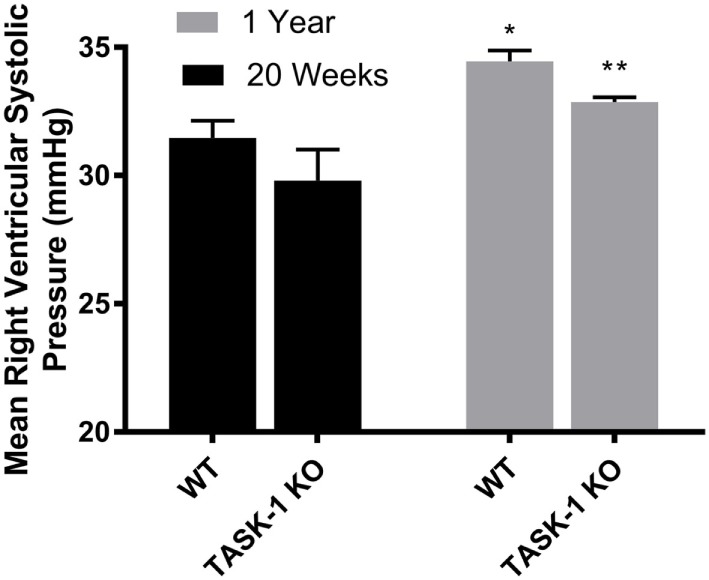
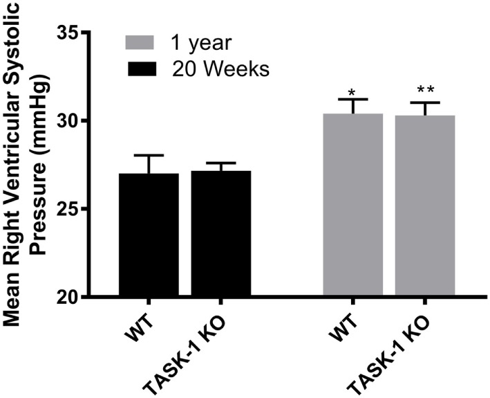
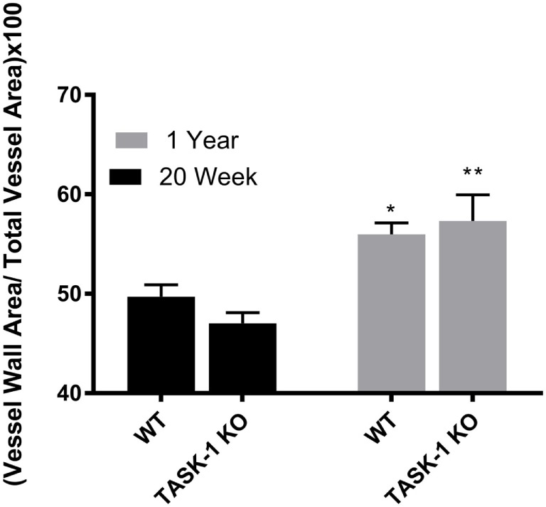
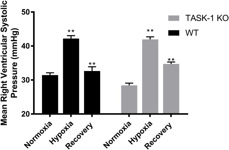
Methods: We measured right ventricular systolic pressure (RVSP) and vascular remodeling in male and female C57BL/6 WT and TASK-1-/- mice at separate time points: 20-24 weeks and 1 year of age. Additionally, we measured RVSP and vascular remodeling in TASK-1-/- and wild-type mice between 13 and 16 weeks of age exposed to 10% hypoxia for 3 weeks followed by recovery to room air conditions for an additional 6 weeks.

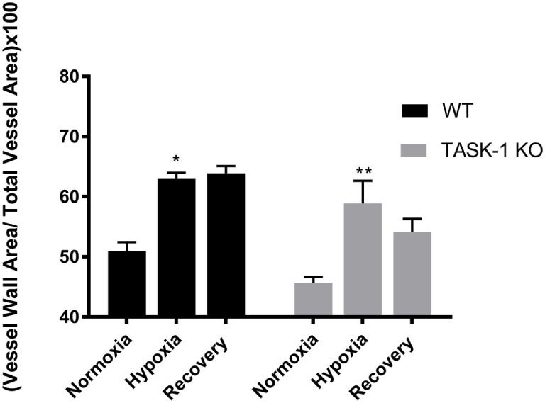
Results: RVSP was similar between WT and TASK-/- mice. Male and female WT and TASK-1-/- mice all demonstrated age-related increases in RVSP, which correlated to age-related vascular remodeling in male mice but not in female mice. Male TASK-1-/- and WT mice exposed to chronic hypoxia demonstrated increased RVSP, which decreased following room air recovery. WT and TASK-1-/- male mice demonstrated vascular remodeling upon exposure to hypoxia that persisted in room air recovery.

Conclusion: Female and male TASK-1-/- mice do not develop hemodynamic or vascular evidence for PH, but RVSP rises in an age-dependent manner independent of genotype. TASK-1-/- and WT male mice develop hypoxia-induced elevations in RVSP that decrease to baseline after recovery in room air. TASK-1-/- and WT male mice demonstrate vascular remodeling after exposure to hypoxia that persists despite recovery to room air conditions and does not correlate with RVSP normalization.

Keywords: KCNK3; TASK-1; potassium channels; pulmonary hypertension; right ventricular systolic pressures.


Figures

Figure 1
Figure 1
Right ventricular systolic pressures (RVSP) at 20 weeks and 1 year in male TASK-1−/− and wild-type C57BL/6 mice. Wild-type male mice demonstrated an age-related increase in RVSP between 20 weeks and 1 year of age (*p < 0.05, n = 5–8). TASK-1−/− male mice also demonstrated an age-related increase in RVSP between 20 weeks and 1 year of age (**p < 0.01, n = 5–8 each group).
Figure 2
Figure 2
Right ventricular systolic pressures (RVSPs) at 20 weeks and 1 year in female TASK-1−/− and wild-type C57BL/6 mice. Wild-type female mice wild-type demonstrated an age-related increase in RVSP between 20 weeks and 1 year of age (*p < 0.05, n = 5–8). TASK-1−/− female mice also demonstrated an age-related increase in RVSP between 20 weeks and 1 year of age (**p < 0.01, n = 5–8 each group).
Figure 3
Figure 3
Ratio of medial wall area: total vessel wall area as an index of vessel wall thickness in male TASK-1−/− and wild-type C57BL/6 male mice at 20 weeks and 1 year. While there was no difference in wall thickness index between genotypes, wild-type male mice wild-type demonstrated an age-related increase in wall thickness between 20 weeks and 1 year of age (*p < 0.05, n = 5–8). TASK-1−/− male mice also demonstrated an age-related increase in right ventricular systolic pressure between 20 weeks and 1 year of age (**p < 0.01, n = 5–8 each group).
Figure 4
Figure 4
Ratio of medial wall area: total vessel wall area as an index of vessel wall thickness in female TASK-1−/− and wild-type C57BL/6 mice at 20 weeks and 1 year. Female TASK-1−/− mice demonstrated a non-significant trend (p = 0.07, n = 6–7 animals per group) in decreased wall thickness compared to female wild-type mice at both 20 weeks and 1 year. There was no measurable age-related increase in wall thickness index in either genotype.
Figure 5
Figure 5
Adult male TASK-1−/− and wild-type mice (C57Bl/6) right ventricular systolic pressures (RVSP) in room air, 10% hypoxia for 3 weeks, then recovery to room air. Wild-type male mice exposed to hypoxia had a significant increase in RVSP hypoxia (*p < 0.05, n = 5) from baseline. Hypoxia-induced elevation in RVSP significantly decreased in recovery to room air (**p < 0.001, n = 6). TASK-1−/− male mice also demonstrated a similar significant increase in RVSP hypoxia (*p < 0.05, n = 6) and that significantly decreased in recovery (**p < 0.01, n = 5). There was no difference between wild-type and TASK-1−/− mice in the degree of increase in RVSP after exposure to hypoxia or the recovery decrease in RVSP.
Figure 6
Figure 6
Adult male TASK-1−/− (KO) and wild-type mice (C57Bl/6) index of wall thickness in room air, 10% hypoxia for 3 weeks, then recovery to room air. Both wild-type (n = 5) and TASK-1−/− (n = 5) animals in hypoxic conditions had increased index of wall thickness compared to baseline normoxia without any difference between genotypes (*p < 0.05, **p < 0.01, wild-type and TASK-1−/−, respectively.) The index of wall thickness remained persistently elevated even in recovery in both genotypes.

References

    1. Galiè N, Humbert M, Vachiery J, Gibbs S, Lang I, Torbicki A, et al. 2015 ESC/ERS Guidelines for the diagnosis and treatment of pulmonary hypertension: the joint task force for the diagnosis and treatment of pulmonary hypertension of the European Society of Cardiology (ESC) and the European Respiratory Society (ERS): endorsed by: Association for European Paediatric and Congenital Cardiology (AEPC), International Society for Heart and Lung Transplantation (ISHLT). Eur Heart J (2016) 37(1):67–119.10.1093/eurheartj/ehv317 - DOI - PubMed
    1. Schermuly RT, Ghofrani HA, Wilkins MR, Grimminger F. Mechanisms of disease: pulmonary arterial hypertension. Nat Rev Cardiol (2011) 8(8):443–55.10.1038/nrcardio.2011.87 - DOI - PMC - PubMed
    1. McLaughlin VV, Archer SL, Badesch DB, Barst RJ, Farber HW, Lindner JR, et al. ACCF/AHA 2009 expert consensus document on pulmonary hypertension. J Am Coll Cardiol (2009) 53(17):1573–619.10.1016/j.jacc.2009.01.004 - DOI - PubMed
    1. Sutendra G, Michelakis ED. Pulmonary arterial hypertension: challenges in translational research and a vision for change. Sci Transl Med (2013) 5(208):1–14.10.1126/scitranslmed.3005428 - DOI - PubMed
    1. Olschewski A. Targeting TASK-1 channels as a therapeutic approach. In: Yuan JJ, Ward J, editors. Advances in Experimental Medicine and Biology. (Vol. 661), Totowa, NJ: Humana Press; (2009). p. 459–73. - PubMed

LinkOut - more resources