

Paget's Breast Disease: A Case Report and Review of the Literature
- PMID: 29109950
- PMCID: PMC5660109
- DOI: 10.3389/fsurg.2017.00051
Paget's Breast Disease: A Case Report and Review of the Literature
Abstract
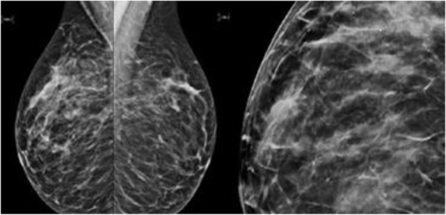
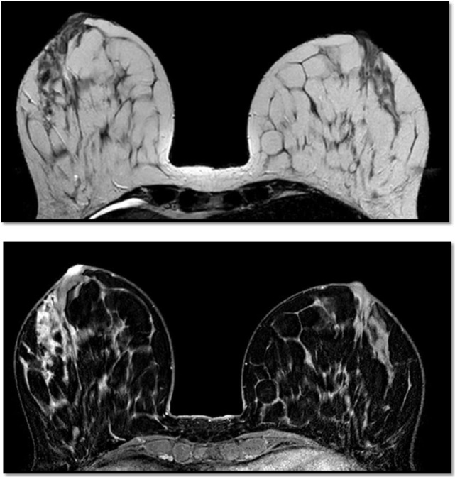
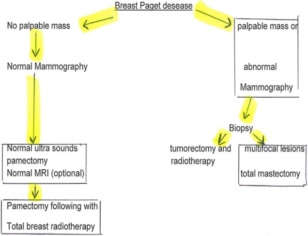
Paget's disease of the breast is a rare cancer. This typical clinical case illustrates the different epidemiological, clinical, histological, therapeutic, and evolving aspects of the disease. We report a case of Paget's disease in a 43-year-old woman who presented eczema of the nipple. Mammography and ultrasounds did reveal a lesion in situ. The patient was scheduled for mastectomy and sentinel node biopsy. She had chosen a radical bilateral surgery. The histological diagnosis was Paget's disease of the breast with a carcinoma in situ. There was no metastasis in either of the sentinel nodes. Paget's disease must be considered with the presence of a persistent eczematous involvement of the nipple, which does not respond to local treatment. Ultrasounds, mammography, and magnetic resonance imaging can allow searching an underlying cancer and guiding the surgical management. There is no evidence at this time that one of the two surgical techniques (conservative or mastectomy) would improve survival. The prognosis depends on the presence of a palpable mass and the invasiveness of the cancer.
Keywords: biopsy; breast Paget’s disease; female breast cancer; imagery; sentinel lymph node biopsy; surgery.
Figures


References
-
- Alvero R. Paget’s disease of the breast. Ferri’s Clinical Advisor 2017. Philadelphia: Elsevier Health Sciences; (2017). 915 p.
-
- Cancer League of Switzerland, 2009–2013. (2016).
-
- Mkhinini I, Fatnassi R, Saidi W, Mansouri W, Rebhi I, Kraiem S, et al. Paget disease of the nipple, Female Imaging EM Consult Maternity Ward, Ibn-El Jazzar Hospital, 3140 Kairouan, Tunisia. (2016).
-
- Vercambre-Darras S, Bertrand M, Daussay D, Mortier L. Paget disease. Dermatology (2012) 7(2):1–9.
Publication types
LinkOut - more resources
Full Text Sources
Other Literature Sources