Review
. 2018 Jan 15;596(2):145-162.
doi: 10.1113/JP273463. Epub 2017 Dec 4.

Studies of cortical connectivity using optical circuit mapping methods

Affiliations
Review

Studies of cortical connectivity using optical circuit mapping methods

Paul G Anastasiades et al. J Physiol. .

Abstract

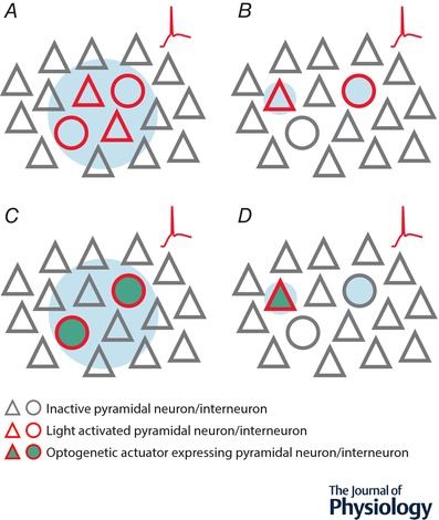
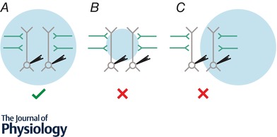
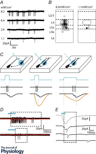
An important consideration when probing the function of any neuron is to uncover the source of synaptic input onto the cell, its intrinsic physiology and efferent targets. Over the years, electrophysiological approaches have generated considerable insight into these properties in a variety of cortical neuronal subtypes and circuits. However, as researchers explore neuronal function in greater detail, they are increasingly turning to optical techniques to bridge the gap between local network interactions and behaviour. The application of optical methods has increased dramatically over the past decade, spurred on by the optogenetic revolution. In this review, we provide an account of recent innovations, providing researchers with a primer detailing circuit mapping strategies in the cerebral cortex. We will focus on technical aspects of performing neurotransmitter uncaging and channelrhodopsin-assisted circuit mapping, with the aim of identifying common pitfalls that can negatively influence the collection of reliable data.

Keywords: cerebral cortex; glutamate uncaging; neural circuits; optogenetics.


Figures

Figure 1
Figure 1. Overview of methods to optically stimulate neuronal populations
A, single‐photon uncaging indiscriminately activates multiple populations of neurons within the local network. Firing (red trace) is induced in all neurons within the spatial extent of the light beam. B, two‐photon uncaging selectively activates individual neurons with single cell resolution. Firing (red trace) can be induced in target cells, but not adjacent neurons. C, single‐photon optogenetics selectively activates multiple neurons within the local network. Firing (red trace) is induced in all neurons within the spatial extent of the light beam so long as they express the construct encoding the optogenetic actuator. D, two‐photon optogenetics selectively activates individual neurons with single cell resolution. Firing (red trace) can be induced in the target cell, but not adjacent neurons, so long as target neurons express the construct encoding the optogenetic actuator.
Figure 2
Figure 2. Strategy to examine long‐range inputs with wide‐field optogenetics
A, presynaptic axons (green) expressing the optogenetic construct are present within cortical network and can be selectively activated by light. Wide‐field illumination must be correctly aligned so the light beam covers the entire extent of the dendritic arbour of both recorded neurons. Input from the presynaptic region expressing the optogenetic actuator can be reliably compared at the two postsynaptic neurons. B, wide‐field illumination is too focused so the light beam only covers a portion of the dendritic arbour of the recorded neurons. Inputs that synapse onto dendrites outside the extent of the light beam are not activated, so total input from the presynaptic region expressing the optogenetic actuator cannot be reliably compared at the two postsynaptic neurons. C, wide‐field illumination is incorrectly aligned as the light beam only covers the dendritic arbour of one of the recorded neurons. Inputs that synapse onto dendrites outside the extent of the light beam are not activated, so inputs onto the leftmost cell cannot be reliably measured.
Figure 3
Figure 3. Optimising LSPS laser calibration and event detection
A, loose cell‐attached recordings of action potentials (APs) evoked by glutamate uncaging in a layer 5a pyramidal cell in primary somatosensory cortex at postnatal day 8 (P8). The laser was fired (1 Hz) across a 50 μm resolution grid that covered the depth of a cortical column at this age. Decreasing laser intensity reduced the number of sites at which APs were elicited. B, average maps showing the distribution of points at which APs can be evoked in a recorded layer 5a pyramidal cell (triangle). At high laser power (left) APs can be evoked across layers 4 to 5b as well as at a single point in layer 2/3. Using a low laser power (right) confined action potential generation to sites in the immediate layer 5a, providing good spatial resolution. C, identifying light‐evoked synaptic responses is straight forward for distal presynaptic neurons (left). However targeting presynaptic neurons close to the recorded cell (middle) invariably results in a direct glutamate response (orange line, bottom traces) with an onset locked to the start of the laser pulse. Large direct responses can obscure evoked synaptic responses. Using a long duration, low intensity laser pulse (right) leads to a slower direct glutamate response from which synaptic responses are more readily extracted (see E). D, action potentials evoked from the same neuron upon laser stimulation across the entire depth of the cortex at the two laser intensities shown in B. The monosynaptic event windows (dashed box) begins at the earliest spike and ends 100 ms after the last spike is detected. Top trace recorded at 8.2 mW cm−2, bottom trace, 1.1 mW cm−2. E, example traces recorded from a layer 5a fast spiking (FS) interneuron: top trace, direct glutamate response evoked when the laser was fired at the cell body of the FS interneuron. Despite the careful calibration and slow laser pulse no excitatory postsynaptic current (EPSC) could be extracted from this single spot. Second trace, EPSCs are evoked as a delay from the laser onset and can be extracted from the low amplitude direct response. Third trace, an EPSC evoked from a pyramidal cell distal from the dendritic arbour of the FS cell. Bottom trace, a spontaneous or polysynaptic EPSC that falls outside of the monosynaptic event detection window.

References

    1. Adesnik H & Scanziani M (2010). Lateral competition for cortical space by layer‐specific horizontal circuits. Nature 464, 1155–1160. - PMC - PubMed
    1. Anastasiades PG & Butt SJ (2011). Decoding the transcriptional basis for GABAergic interneuron diversity in the mouse neocortex. Eur J Neurosci 34, 1542–1552. - PubMed
    1. Anastasiades PG & Butt SJ (2012). A role for silent synapses in the development of the pathway from layer 2/3 to 5 pyramidal cells in the neocortex. J Neurosci 32, 13085–13099. - PMC - PubMed
    1. Anastasiades PG, Marques‐Smith A, Lyngholm D, Lickiss T, Raffiq S, Katzel D, Miesenbock G & Butt SJ (2016). GABAergic interneurons form transient layer‐specific circuits in early postnatal neocortex. Nat Commun 7, 10584. - PMC - PubMed
    1. Anderson CT, Sheets PL, Kiritani T & Shepherd GM (2010). Sublayer‐specific microcircuits of corticospinal and corticostriatal neurons in motor cortex. Nat Neurosci 13, 739–744. - PMC - PubMed

Publication types