. 2018 Jun;141(6):2142-2155.e5.
doi: 10.1016/j.jaci.2017.08.040. Epub 2017 Oct 27.

Ruxolitinib partially reverses functional natural killer cell deficiency in patients with signal transducer and activator of transcription 1 (STAT1) gain-of-function mutations

Affiliations

Ruxolitinib partially reverses functional natural killer cell deficiency in patients with signal transducer and activator of transcription 1 (STAT1) gain-of-function mutations

Alexander Vargas-Hernández et al. J Allergy Clin Immunol. 2018 Jun.

Abstract

Background: Natural killer (NK) cells are critical innate effector cells whose development is dependent on the Janus kinase-signal transducer and activator of transcription (STAT) pathway. NK cell deficiency can result in severe or refractory viral infections. Patients with STAT1 gain-of-function (GOF) mutations have increased viral susceptibility.

Objective: We sought to investigate NK cell function in patients with STAT1 GOF mutations.

Methods: NK cell phenotype and function were determined in 16 patients with STAT1 GOF mutations. NK cell lines expressing patients' mutations were generated with clustered regularly interspaced short palindromic repeats (CRISPR-Cas9)-mediated gene editing. NK cells from patients with STAT1 GOF mutations were treated in vitro with ruxolitinib.

Results: Peripheral blood NK cells from patients with STAT1 GOF mutations had impaired terminal maturation. Specifically, patients with STAT1 GOF mutations have immature CD56dim NK cells with decreased expression of CD16, perforin, CD57, and impaired cytolytic function. STAT1 phosphorylation was increased, but STAT5 was aberrantly phosphorylated in response to IL-2 stimulation. Upstream inhibition of STAT1 signaling with the small-molecule Janus kinase 1/2 inhibitor ruxolitinib in vitro and in vivo restored perforin expression in CD56dim NK cells and partially restored NK cell cytotoxic function.

Conclusions: Properly regulated STAT1 signaling is critical for NK cell maturation and function. Modulation of increased STAT1 phosphorylation with ruxolitinib is an important option for therapeutic intervention in patients with STAT1 GOF mutations.

Keywords: Janus kinase inhibition; Signal transducer and activator of transcription 1, gain of function; natural killer cell deficiency; natural killer cell maturation; perforin; ruxolitinib.


Conflict of interest statement

Disclosure of conflicts of interest:

The authors declare no conflict of interest

Figures
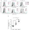
FIG 1
FIG 1
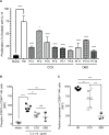
STAT1 GOF mutations lead to delayed dephosphorylation in NK cells. Dephosphorylation in NK cells stimulated with IFN-α for the indicated periods in healthy donors, A, patients with mutations into the Coiled-coil domain (CCD; patients 2, 3, 4, 5, 6, 7, and 8) or DNA-binding domain (DBD; patients 9, 13, and 15). B, levels of STAT1-activated after 120 minutes of stimulation. HD, healthy donor; Pt; patient. The statistical significance was analyzed using the Ordinary one-way ANOVA test ***p<0.001, ****p<0.0001.
FIG 2
FIG 2
Impaired NK cell cytotoxic capacity in patients with STAT1 GOF mutations. NK cell cytotoxicity was measured by Cr release in the presence or absence of 1000 U/mL of IL-2. A, patients with Coiled-coil domain (CCD) mutations; B, patients with DNA-binding domain (DBD) mutations. HD, healthy donor; Pt; patient.
FIG 3
FIG 3
CD56dim NK cells have an immature phenotype in patients with STAT1 GOF mutations. NK cell phenotype from healthy donors and STAT1 GOF patients were analyzed by flow cytometry. A, NK cell subsets were defined as CD56brightCD3 cells or CD56dimCD3. B, representative graph shows the frequencies of CD56brightCD3 and CD56dimCD3 NK cells subsets. C, representative graphs shows mean fluorescence intensity (MFI) for each NK cell receptor expressed by CD56dim NK cells. Each blue circle represents a healthy donor and a red box represents a STAT1 GOF patient. Horizontal bars represent means and the vertical bars indicate the standard deviation. The statistical significance was analyzed using the unpaired Student’s t-test. Results are representative of 3 independent experiments. **p<0.01, ***p<0.001, ****p<0.0001.
FIG 4
FIG 4
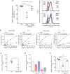
STAT1 GOF mutations affect the cytotoxic capacity and perforin expression in NK cell lines. DNA-binding domain mutations E353K and T385M were expressed in the YTS cell line using CRISPR-Cas9 gene editing. A, STAT1 hyper-phosphorylation in response to IFN-α was measured by intracellular flow cytometry using pY701-STAT1 antibody. B, the phenotype of YTS STAT1 GOF cell lines was measured by flow cytometry. C, NK cell cytotoxicity was analyzed against K562 target cells using standard Cr release protocol. D, STAT1 hyper753 phosphorylation in response to IFN-α; perforin, NKG2A, and CD94 expression, and cytotoxic capacity measured in NK92 STAT1-M202I mutant cell line. MFI, Mean fluorescence intensity; WT, wild type. Results are representative of 3 independent experiments. * p<0.05.
FIG 5
FIG 5
Decreased STAT5 activation in CD56dim NK cells from STAT1 GOF patients. A, NK cell proliferation from healthy donors and STAT1 GOF patients in presence of IL-15 (5 ng/mL). B, phospho-STAT5 levels in CD56dim NK cells stimulated with IL-2 (10 ng/ml) for 30 minutes. Each point represents the mean fluorescence intensity for each individual healthy donor or patients. C, perforin expression levels in CD56dim NK cells from patients with CCD or DBD mutations. Statistical significance was analyzed using the Ordinary one-way ANOVA test. * p<0.05, **p<0.01, ***p<0.001, ****p<0.0001. HD, healthy donors; MFI, Mean fluorescence intensity; CCD, Coiled-coil domain (patients 2, 4, 5, 6, 7, and 8); DBD,DNA-binding domain (patients 11, 15, and 16). Results are representative of 2 independent experiments for each patient.
FIG 6
FIG 6
Ruxolitinib restores perforin expression in primary NK cells. A, percentages of CD56dimPerforin+ NK cells and perforin expression on CD56dim NK cell subset pre- and post-treatment. B, natural cytotoxicity was performed with Cr release protocol with freshly isolated PBMC from patient 9, and 11 and a respective healthy donor and was tested against K562 target cells in the presence or absence of 1000 U/mL of IL-2. STAT5 activation in CD56dim NK cells after stimulation with IL-2, D, patients 3, 9, and 11 (pre-treatment) and D, in CD56dim NK cells from patient 9 and 11post-treatment. E, PBMCs from Pt 13 were incubated in vitro for 48 hours with or without 1 µM of ruxolitinib and the perforin expression was measured in CD56dim NK cells. The statistical significance was analyzed using the Ordinary one-way ANOVA test * p<0.05, **p<0.01, ***p<0.001.

References

    1. Cooper MA, Fehniger TA, Caligiuri MA. The biology of human natural killer-cell subsets. Trends Immunol. 2001;22:633–40. - PubMed
    1. Biron CA, Nguyen KB, Pien GC, Cousens LP, Salazar-Mather TP. Natural killer cells in antiviral defense: function and regulation by innate cytokines. Annu Rev Immunol. 1999;17:189–220. - PubMed
    1. Caligiuri MA. Human natural killer cells. Blood. 2008;112:461–9. - PMC - PubMed
    1. Cooper MA, Fehniger TA, Turner SC, Chen KS, Ghaheri BA, Ghayur T, et al. Human natural killer cells: a unique innate immunoregulatory role for the CD56(bright) subset. Blood. 2001;97:3146–51. - PubMed
    1. Poli A, Michel T, Theresine M, Andres E, Hentges F, Zimmer J. CD56bright natural killer (NK) cells: an important NK cell subset. Immunology. 2009;126:458–65. - PMC - PubMed

Publication types