. 2018 Mar-Apr;20(2):166-172.
doi: 10.4103/aja.aja_49_17.

Lipoxin A4 improves erectile dysfunction in rats with type I diabetes by inhibiting oxidative stress and corporal fibrosis

Affiliations

Lipoxin A4 improves erectile dysfunction in rats with type I diabetes by inhibiting oxidative stress and corporal fibrosis

Kai Cui et al. Asian J Androl. 2018 Mar-Apr.

Abstract

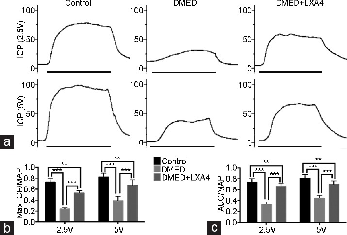
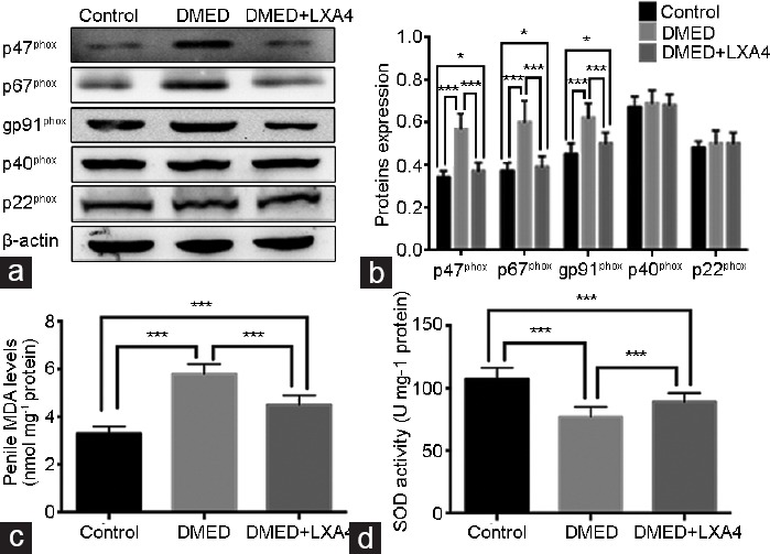
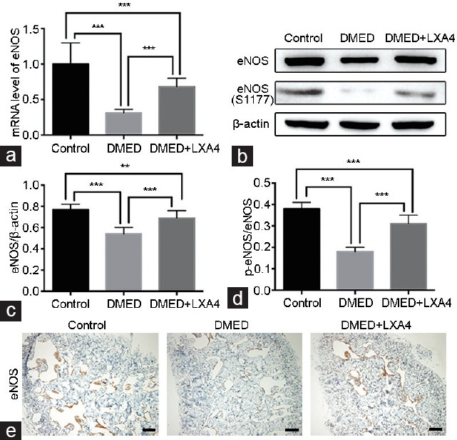
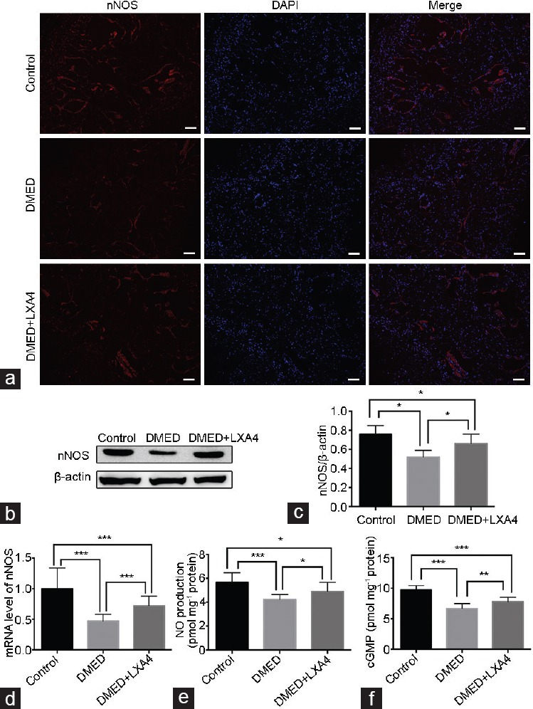
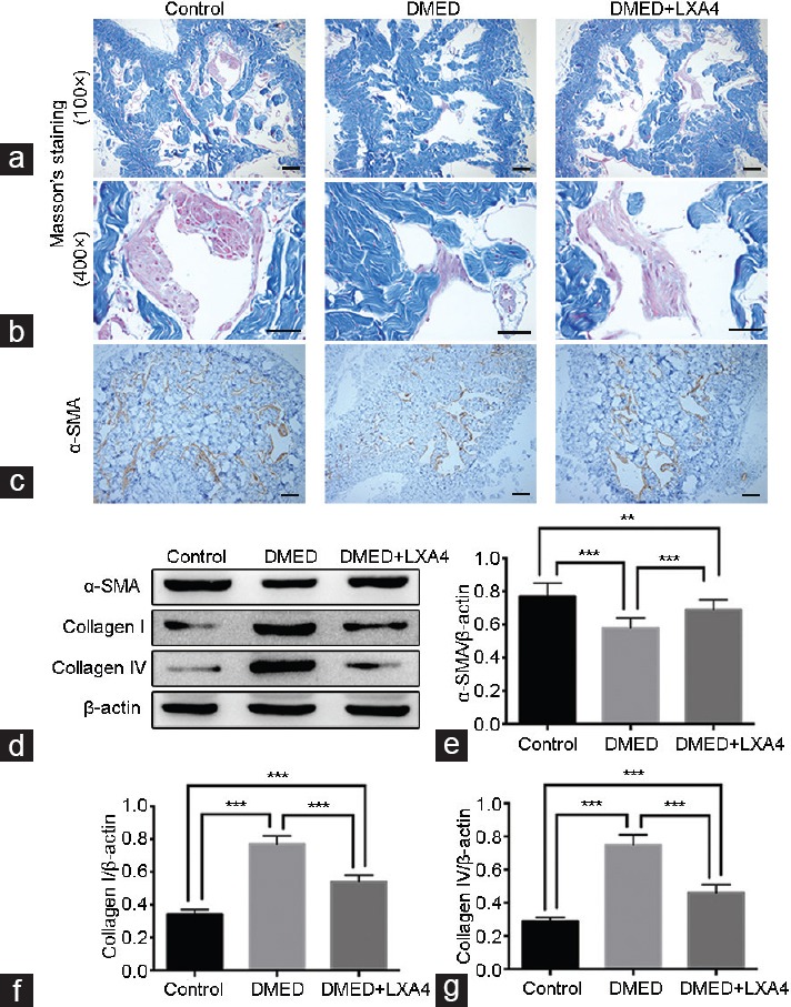
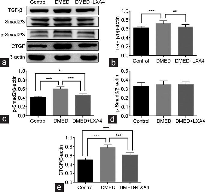
Previous studies have shown that oxidative stress and corporal fibrosis in penile tissues of rats were key pathological factors of erectile dysfunction induced by diabetic mellitus (DMED). Lipoxin A4 (LXA4) was reported to inhibit oxidative stress and fibrosis diseases, while whether it could exert a protective role on erectile function was not clear. Type I diabetic mellitus (DM) was induced in thirty male 10-week-old Sprague-Dawley rats using streptozotocin. Ten weeks later, twenty-two rats with DMED confirmed by an apomorphine test were divided into two groups: the DMED group (n = 11) and the DMED + LXA4 group (n = 11; LXA4 injection daily for 4 weeks). In addition, another ten age-matched rats formed the Control group. We found that erectile function was significantly impaired in the DMED group compared with the Control group, but was improved in the DMED + LXA4 group. Similarly, the over-activated oxidative stress and impaired endothelial function in the DMED group were both improved in the DMED + LXA4 group. Moreover, the DMED group showed serious corporal fibrosis, which was also inhibited by the treatment of LXA4 in the DMED + LXA4 group. Taken together, LXA4 could exert an inhibition role on oxidative stress and fibrosis to improve DMED effectively.

Keywords: diabetic mellitus; erectile dysfunction; fibrosis; oxidative stress.


Figures

Figure 1
Figure 1
Treatment of LXA4 improves erectile dysfunction elicited by electrical stimulation of the cavernous nerve. (a) Representative ICP tracings were measured through stimulation of 2.5 and 5 V for 1 min, respectively. Ratios of max ICP and AUC to MAP of all the three groups were presented through bar graphs: (b) for max ICP/MAP and (c) for AUC/MAP. Data are expressed as mean ± s.e.m. (ncontrol = 10, nDMED = 11, and nDMED + LXA4 = 11). **P < 0.01 and ***P < 0.001 when comparing two groups. DMED: diabetes mellitus-induced erectile dysfunction; LXA4: Lipoxin A4; ICP: intracavernous pressure; AUC: area under the curve; MAP: mean arterial pressure. Scale bars = 1 min; s.e.m.: standard error of the mean.
Figure 2
Figure 2
Treatment of LXA4 inhibits oxidative stress in corpus cavernous of DMED rats. (a) Representative Western blot results for the p22phox, p47phox, gp91phox, p40phox, and p67phox in rats of all the three groups. (b) Expressions of p22phox, p47phox, gp91phox, p40phox, and p67phox with β-actin as the loading control in all the three groups were presented through bar graphs. (c) MDA levels determined by the ELISA method in all the three groups. (d) SOD activities determined by the ELISA method in all the three groups. Data are expressed as mean ± s.e.m. (ncontrol = 10, nDMED = 11, and nDMED + LXA4 = 11). *P < 0.05 and ***P < 0.001 when comparing two groups. DMED: diabetes mellitus-induced erectile dysfunction; LXA4: Lipoxin A4; ELISA: enzyme-linked immunosorbent assay; SOD: superoxide dismutase; MDA: malondialdehyde; s.e.m.: standard error of the mean.
Figure 3
Figure 3
Treatment of LXA4 increases eNOS activation in corpus cavernous of DMED rats. (a) eNOS expression level with β-actin as the loading control in penile tissues of all groups by real-time RT-PCR using the 2-ΔΔCt method. (b) Representative Western blot results for eNOS and p-eNOS (S1177) of rats in the Control, DMED, and DMED + LXA4 groups after 4 weeks' treatment. Expressions of eNOS (c) with β-actin as the loading control and its phosphorylation level at the S177 site (d) in all the three groups were presented through bar graphs. (e) Immunohistochemical staining with antibody to eNOS (×100). Scale bars = 100 μm. Data are expressed as mean ± s.e.m. (ncontrol = 10, nDMED = 11, and nDMED + LXA4 = 11). **P < 0.01 and ***P < 0.001 when comparing two groups. DMED: diabetes mellitus-induced erectile dysfunction; LXA4: Lipoxin A4; RT-PCR: reverse transcriptase-polymerase chain reaction; eNOS: endothelial nitric oxide synthase; s.e.m.: standard error of the mean.
Figure 4
Figure 4
Treatment of LXA4 increases nNOS expression, thus activates the NO/cGMP signaling pathway. (a) Immunofluorescence results of nNOS in all the three groups after 4 weeks' treatment (×100). Scale bars = 100 μm. (b) Representative Western blot results for nNOS of rats in the Control, DMED, and DMED + LXA4 groups after 4 weeks' treatment. (c) Expressions of nNOS with β-actin as the loading control in all the three groups were presented through bar graphs. (d) nNOS expression level with β-actin as the loading control in penile tissues of all groups by real-time RT-PCR using the 2-ΔΔCt method. (e) The NO concentrations in all the three groups determined by the ELISA method. (f) The cGMP concentrations in all the three groups determined by the ELISA method. Data are expressed as mean ± s.e.m. (ncontrol = 10, nDMED = 11, and nDMED + LXA4 = 11). *P < 0.05, **P < 0.01, and ***P < 0.001 when comparing two groups. DMED: diabetes mellitus-induced erectile dysfunction; LXA4: Lipoxin A4; nNOS: neuronal nitric oxide synthase; NO: nitric oxide; cGMP: cyclic guanosine monophosphate; RT-PCR: reverse transcriptase-polymerase chain reaction; s.e.m.: standard error of the mean.
Figure 5
Figure 5
Treatment of LXA4 inhibits corporal fibrosis in corpus cavernous of DMED rats. Masson's trichrome staining of magnification ×100 (a) and magnification ×400 (b) was performed to assess the corporal fibrosis level in rat cavernous of all the three groups after 4 weeks' treatment (the area of smooth muscle is represented by red stain and the area of collagen by blue stain). Scale bars = 100 μm. (c) Immunohistochemistry results of α-SMA (×100) in all the three groups. Scale bars = 100 μm. (d) Representative Western blot results for a-SMA, Collagen I, and Collagen IV in all the three groups. Expressions of (e) α-SMA, (f) Collagen I, and (g) Collagen IV with β-actin as the loading control in all the three groups were presented through bar graphs. Data are expressed as mean ± s.e.m. (ncontrol = 10, nDMED = 11, and nDMED + LXA4 = 11). **P < 0.01 and ***P < 0.001 when comparing two groups. DMED: diabetes mellitus-induced erectile dysfunction; LXA4: Lipoxin A4; α-SMA: α-smooth muscle actin; s.e.m.: standard error of the mean.
Figure 6
Figure 6
Treatment of LXA4 inhibits the TGF-β1/Smad/CTGF signaling pathway activity. (a) Representative Western blot results for TGF-β1, Smad2/3, p-Smad2/3, and CTGF in all the three groups after 4 weeks' treatment. Expressions of (b) TGF-β1, (c) p-Smad2, (d) p-Smad3, and (e) CTGF with β-actin as the loading control in all the three groups were presented through bar graphs. Data are expressed as mean ± s.e.m. (ncontrol = 10, nDMED = 11, and nDMED + LXA4 = 11). *P < 0.05, **P < 0.01, and ***P < 0.001 when comparing two groups. DMED: diabetes mellitus-induced erectile dysfunction; LXA4: Lipoxin A4; TGF-β1: transforming growth factor-beta 1; CTGF: connective tissue growth factor; s.e.m.: standard error of the mean.

References

    1. Shamloul R, Ghanem H. Erectile dysfunction. Lancet. 2013;381:153–65. - PubMed
    1. Shiri R, Koskimaki J, Hakkinen J, Tammela TL, Huhtala H, et al. Effects of age, comorbidity and lifestyle factors on erectile function: Tampere Ageing Male Urological Study (TAMUS) Eur Urol. 2004;45:628–33. - PubMed
    1. Feldman HA, Goldstein I, Hatzichristou DG, Krane RJ, McKinlay JB. Impotence and its medical and psychosocial correlates: results of the Massachusetts Male Aging Study. J Urol. 1994;151:54–61. - PubMed
    1. Johannes CB, Araujo AB, Feldman HA, Derby CA, Kleinman KP, et al. Incidence of erectile dysfunction in men 40 to 69 years old: longitudinal results from the Massachusetts Male Aging Study. J Urol. 2000;163:460–3. - PubMed
    1. Andersen I, Heitmann BL, Wagner G. Obesity and sexual dysfunction in younger Danish men. J Sex Med. 2008;5:2053–60. - PubMed

MeSH terms