. 2017 Dec 14;60(23):9545-9564.
doi: 10.1021/acs.jmedchem.7b00861. Epub 2017 Nov 21.

Structure-Affinity Relationships and Structure-Kinetic Relationships of 1,2-Diarylimidazol-4-carboxamide Derivatives as Human Cannabinoid 1 Receptor Antagonists

Affiliations

Structure-Affinity Relationships and Structure-Kinetic Relationships of 1,2-Diarylimidazol-4-carboxamide Derivatives as Human Cannabinoid 1 Receptor Antagonists

Lizi Xia et al. J Med Chem. .

Abstract

We report on the synthesis and biological evaluation of a series of 1,2-diarylimidazol-4-carboxamide derivatives developed as CB1 receptor antagonists. These were evaluated in a radioligand displacement binding assay, a [35S]GTPγS binding assay, and in a competition association assay that enables the relatively fast kinetic screening of multiple compounds. The compounds show high affinities and a diverse range of kinetic profiles at the CB1 receptor and their structure-kinetic relationships (SKRs) were established. Using the recently resolved hCB1 receptor crystal structures, we also performed a modeling study that sheds light on the crucial interactions for both the affinity and dissociation kinetics of this family of ligands. We provide evidence that, next to affinity, additional knowledge of binding kinetics is useful for selecting new hCB1 receptor antagonists in the early phases of drug discovery.


Conflict of interest statement

The authors declare no competing financial interest.

Figures

Figure 1
Figure 1
Chemical structures of (a) rimonabant, (b) taranabant, (c) otenabant, and (d) the scaffold of 1,2-diarylimidazol-4-carboxamides as CB1 receptor antagonists; the R1 substitution is defined as the “left arm” of the scaffold while the R2 substitution defines the “right arm” of the scaffold. The calculations of PSA values are reported in Supporting Information.
Scheme 1
Scheme 1. Synthesis of Antagonists 8a, 8b, and 11ah
Reagents and conditions: (a) EtMgBr, 2,4-diClPhCN, THF, rt, 20 h, 98%; (b) (i) EtO2CC(O)CH(Br)CH3, K2CO3, THF, rt, 66 h, (ii) AcOH, reflux, 1 h, 65%; (c) HBr, AcOH, rt, 15 h, 63%; (d) R1-OH, DEAD, Ph3P, THF, toluene, rt, 15 h, 77%; (e) KOH, EtOH:THF:H2O 2:2:1, 50 °C, 3.5 h, 95%; (f) (i) (COCl)2, DMF cat., CH2Cl2, rt, 90 min, (ii) piperidin-1-amine·HCl, pyridine, CH2Cl2, rt, 2 h, 55% (2 steps); (g) KOH, MeOH:H2O 3:1, reflux, 2 h, 99%; (h) (i) (COCl)2, DMF cat., CH2Cl2, reflux, 2 h, (ii) piperidin-1-amine, NEt3, CH2Cl2, 0 °C to rt, 2 h, 74%; (i) BBr3, CH2Cl2, rt, 1 h, 58%; (j) R1-X, base, CH2Cl2. Corresponding 56–90% R1 substitutions are listed in Table 1.
Scheme 2
Scheme 2. Synthesis of Antagonists 14a14h, 19, (±)-22, (±)-25, and 28
Reagents and conditions: (a) (i) SOCl2, reflux or (COCl)2, DMF cat., CH2Cl2, rt, (ii) R2-NH2, NEt3, CH2Cl2, 17–98% (2 steps), or 2-amino-5-trifluoromethylpyridine, Me3Al, CH2Cl2, rt to 45 °C, 16 h, 64%; (b) BF3·OEt2, Me2S, CH2Cl2, rt, or HBr, AcOH, rt, 20–97%; (c) Et3N, F3CCH2CH2SO2Cl, CH2Cl2, −78 °C, 25–97%; (d) (i) TBDMSCl, Et3N, CH2Cl2, rt, 22 h, (ii) Boc2O, THF, rt, 4 h, 70% (4 steps, a, b, d i, and d ii), (iii) TBAF, THF, rt, 90 min, (iv) F3CCH2CH2SO2Cl, Et3N, CH2Cl2, −78 °C, 3 h, (v) SOCl2, MeOH, 0 °C to rt, 1 h, 56% (3 steps, d iii., d iv, and d v); (e) (i) (COCl)2, DMF cat., CH2Cl2, rt, 2 h, (ii) Cl3CCH2OH, NEt3, CH2Cl2, rt, 3 h, 95% (2 steps, e, b); (f) Zn, AcOH, 3 h; (g) (i) (COCl)2, DMF cat., CH2Cl2, rt, 2 h, (ii) 4-aminocyclohexanol, NaOH, H2O:CH2Cl2 2:1, rt, 2 h, 54% (2 steps, f, g); (h) CH2O, NaBH4, NaBH3CN, CH3CN, H2O, AcOH, rt, 48 h, 32%. Corresponding R2 substitutions are listed in Table 2.
Figure 2
Figure 2
Correlation between the affinities/potencies of the CB1 receptor antagonists measured in a radioligand binding assay (X-axis) and in a GTPγS binding assay (Y-axis) (r2 = 0.49, P = 0.0001). Data taken from Tables 1 and 2
Figure 3
Figure 3
Association and dissociation profile of [3H]CP55940 (2.9 nM) at recombinant hCB1 receptors stably expressed on CHO cell membranes at 30 °C. After 120 min of association, unlabeled rimonabant (10 μM) was added to initiate the dissociation. Association data was fitted in Prism 6 using one-phase exponential association (n = 3, combined and normalized). Dissociation data was fitted using one-phase exponential decay (n = 4, combined and normalized). Data are shown as mean ± SEM from at least three separate experiments each performed in duplicate.
Figure 4
Figure 4
(a) Competition association experiments with [3H]CP55940 binding to recombinant hCB1 receptors stably expressed on CHO cell membranes (30 °C) in the absence or presence of 3.5, 11, and 35 nM of unlabeled CP55940 (n = 3, combined and normalized). (b) Competition association experiments with [3H]CP55940 binding to recombinant hCB1 receptors stably expressed on CHO cell membranes (30 °C) in the absence or presence of 120 nM of unlabeled Rimonabant (n = 6, representative graph). t1 is the radioligand binding at 30 min, while t2 is the radioligand binding at 240 min.
Figure 5
Figure 5
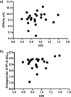
(a) Negative logarithm of the affinities of the hCB1 receptor antagonists used in this study had no obvious linear correlation with their KRI values (r2 = 0.04, P = 0.33). (b) Negative logarithm of [35S]GTPγS IC50 values of the hCB1 receptor antagonists in this study had no obvious linear correlation with their KRI values (r2 = 0.12, P = 0.10).
Figure 6
Figure 6
Competition association experiments with [3H]CP55940 binding to recombinant hCB1 receptors stably expressed on CHO cell membranes (30 °C) in the absence or presence of unlabeled long residence time compound 28 (8.22 nM, red, representative curve) or short residence time compound 11b (12.72 nM, blue, representative curve). Data are shown as mean values from one representative experiment. At least three separate experiments each performed in duplicate.
Figure 7
Figure 7
CP55940-stimulated [35S]GTPγS binding to recombinant hCB1 receptors stably expressed on CHO cell membranes (25 °C) in the absence (black, representative curve) or presence of long-residence-time compound 28 (red, representative curve) or rimonabant (blue, representative curve). Compound 28 or rimonabant was preincubated with the membranes for 1 h prior to the challenge of agonist. [35S]GTPγS was subsequently added and incubated for another 0.5 h. Plates were then filtered and the radioactivity counted. Curves were fitted to a four parameter logistic dose–response equation. Data were normalized according to the maximal response (100%) produced by CP55940. At least three separate experiments each performed in duplicate.
Figure 8
Figure 8
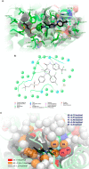
(a) Docking of antagonist 28 into the binding site of the crystal structure of the CB1 receptor (PDB 5TGZ) co-crystallized with 29 (not shown). Compound 28 is represented by black sticks, and residues within 5 Å of 28 are visualized as green sticks. The protein is represented by green ribbons, and relevant binding site confinements are indicated by white-gray (hydrophobic), red (electronegative), and blue (electropositive) layers. Ligand and residues atoms color code: yellow = sulfur, red = oxygen, blue = nitrogen, cyan = fluorine, white = hydrogen. (b) 2-D interaction map of 28 docking into the CB1 receptor co-crystallized with 29 (PDB 5TGZ), demonstrating π–π stacking between imidazole core of 28 and Phe102N-term, 2,4-dichlorophenyl ring and Phe1702.57, and pyridine and His1782.65. (c) Docking of 14f and 28 into the binding site of the crystal structure of the CB1 receptor co-crystallized with 29 (PDB 5TGZ), showing the overlay of numbered consecutively hydration sites of 14f (colored spheres; for color code, see below) calculated by WaterMap. Hydration sites shown as red and orange spheres represent “unstable” water molecules. White spheres symbolize “stable” water molecules, which should not be displaced by 14f or 28. For the key hydration sites (41, 69, 72, 81, 88) surrounding the −F atom of 14f, calculated ΔG values (in kcal/mol) with respect to bulk solvent are shown.

Similar articles

Cited by

References

    1. Maccarrone M.; Bab I.; Bíró T.; Cabral G. A.; Dey S. K.; Di Marzo V.; Konje J. C.; Kunos G.; Mechoulam R.; Pacher P.; Sharkey K. A.; Zimmer A. Endocannabinoid signaling at the periphery: 50 years after THC. Trends Pharmacol. Sci. 2015, 36, 277–296. 10.1016/j.tips.2015.02.008. - DOI - PMC - PubMed
    1. Pertwee R. G.; Howlett A. C.; Abood M. E.; Alexander S. P. H.; Di Marzo V.; Elphick M. R.; Greasley P. J.; Hansen H. S.; Kunos G.; Mackie K.; Mechoulam R.; Ross R. A. International union of basic and clinical pharmacology. LXXIX. Cannabinoid receptors and their ligands: beyond CB1 and CB2. Pharmacol. Rev. 2010, 62, 588–631. 10.1124/pr.110.003004. - DOI - PMC - PubMed
    1. Munro S.; Thomas K. L.; Abu-Shaar M. Molecular characterization of a peripheral receptor for cannabinoids. Nature 1993, 365, 61–65. 10.1038/365061a0. - DOI - PubMed
    1. Abood M.; Barth F.; Bonner T. I.; Cabral G.; Casellas P.; Cravatt B. F.; Devane W. A.; Elphick M. R.; Felder C. C.; Herkenham M.; Howlett A. C.; Kunos G.; Mackie K.; Mechoulam R.; Pertwee R. G.. Cannabinoid receptors: CB1 receptor. In IUPHAR/BPS Guide to Pharmacology; International Union of Basic and Clinical Pharmacology, May 16, 2017; http://www.guidetopharmacology.org/GRAC/ObjectDisplayForward?objectId=56 (accessed July 20, 2017).
    1. Pacher P.; Bátkai S.; Kunos G. The endocannabinoid system as an emerging target of pharmacotherapy. Pharmacol. Rev. 2006, 58, 389–462. 10.1124/pr.58.3.2. - DOI - PMC - PubMed

Publication types

LinkOut - more resources