. 2018 Jan;118(1):43-51.
doi: 10.1038/bjc.2017.374. Epub 2017 Nov 7.

Targeting de novo lipogenesis as a novel approach in anti-cancer therapy

Affiliations

Targeting de novo lipogenesis as a novel approach in anti-cancer therapy

Katharina Stoiber et al. Br J Cancer. 2018 Jan.

Abstract

Background: Although altered membrane physiology has been discussed within the context of cancer, targeting membrane characteristics by drugs being an attractive therapeutic strategy has received little attention so far.

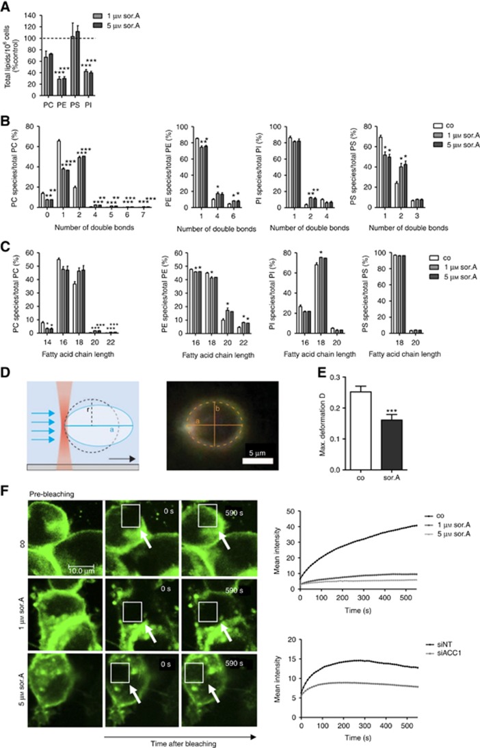
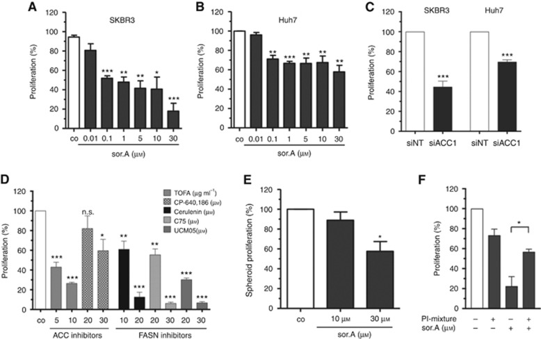
Methods: Various acetyl-CoA carboxylase 1 (ACC1), and fatty acid synthase (FASN) inhibitors (like Soraphen A and Cerulenin) as well as genetic knockdown approaches were employed to study the effects of disturbed phospholipid composition on membrane properties and its functional impact on cancer progression. By using state-of-the-art methodologies such as LC-MS/MS, optical tweezers measurements of giant plasma membrane vesicles and fluorescence recovery after photobleaching analysis, membrane characteristics were examined. Confocal laser scanning microscopy, proximity ligation assays, immunoblotting as well as migration, invasion and proliferation experiments unravelled the functional relevance of membrane properties in vitro and in vivo.

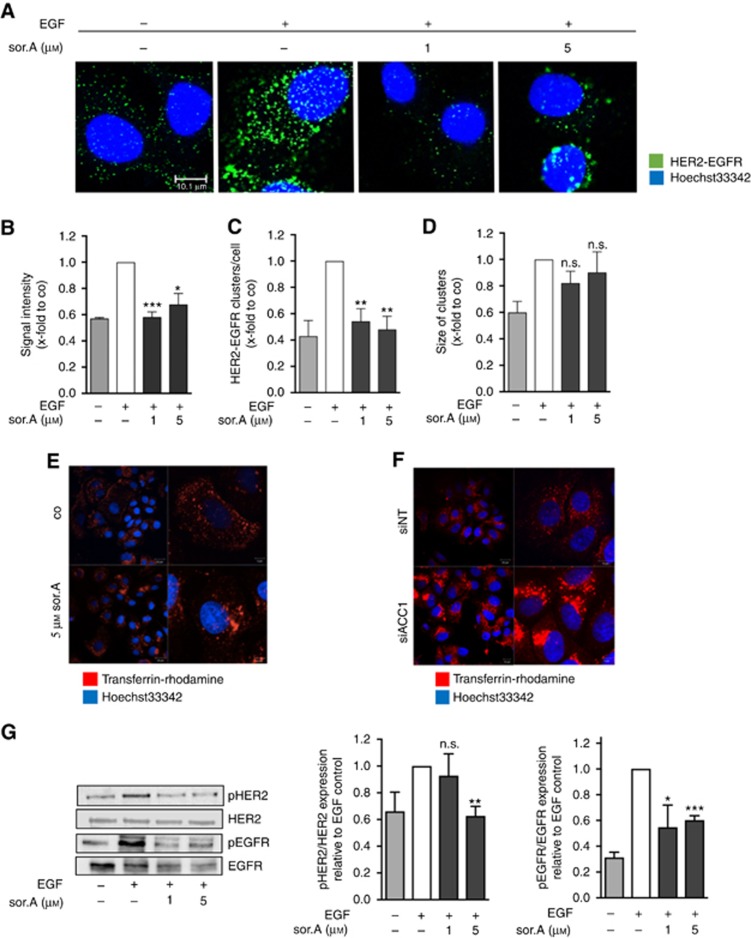
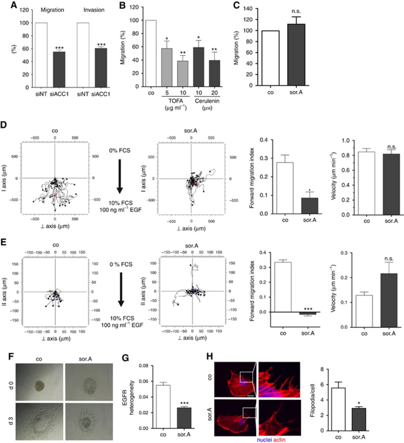
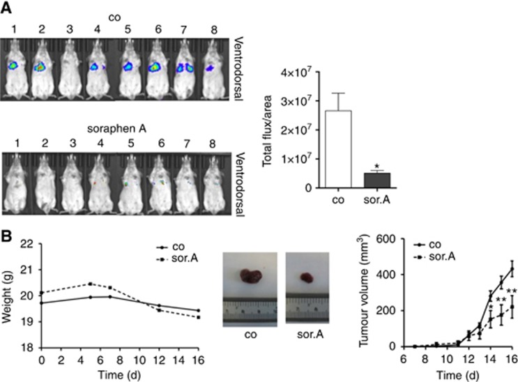
Results: By disturbing the deformability and lateral fluidity of cellular membranes, the dimerisation, localisation and recycling of cancer-relevant transmembrane receptors is compromised. Consequently, impaired activation of growth factor receptor signalling cascades results in abrogated tumour growth and metastasis in different in vitro and in vivo models.

Conclusions: This study highlights the field of membrane properties as a promising druggable cellular target representing an innovative strategy for development of anti-cancer agents.


Conflict of interest statement

The authors declare no conflict of interest.

Figures

Figure 1
Figure 1
Cell mechanics is strongly influenced by ACC1 inhibition. (A) Lipids of soraphen A-treated SKBR3 cells (72 h) were extracted and analysed by liquid chromatography ESI tandem mass spectrometry. Total signal intensities of phospholipid subclasses in soraphen A-treated cells were normalised to control. A value of 100% was assigned to the signal intensities of control cells. (B) Effect of soraphen A on the number of double bonds in fatty acids and (C) the fatty acid chain length of the major phospholipid subclasses phosphatidylcholine (PC), phosphatidylserine (PS), phosphatidylethanolamine (PE) and phosphatidylinositol (PI). Signal intensities are shown relative to the total phospholipid subclass intensity. (D) Experimental setup for vesicle deformation measurements: A single membrane vesicle derived from SKBR3 cells is trapped on one end with a focused laser beam (optical tweezer). Application of flow results in elongation of the vesicle along the flow direction. Right: Dark-field microscopy image of an optically trapped vesicle. The 2D projection of the vesicle is fitted with an ellipse using home-built Matlab and C++ routines. A dashed line indicates an elliptic fit of the vesicle cross-section with half axis a and b. (E) Comparison of the maximum deformation Dmax of vesicles obtained from cells after treatment with soraphen A or control. The error bars indicate the s.e.m. for a sample size of 30 and 40 cells, respectively. (F) Lateral membrane fluidity was assessed by FRAP assay in soraphen A-treated SKBR3 cells and after siACC1 transfection, respectively. Representative images are depicted on the left. White arrows indicate the photobleached area. Right: mean fluorescence recovery after photobleaching is diagrammed over time. Statistical analysis was performed using Student’s t test: *P<0.05, **P<0.01, ***P<0.001.
Figure 2
Figure 2
Impaired dimerisation, recycling and activation of transmembrane receptors. (A) HER2-EGFR dimerisation of SKBR3 cells treated for 72 h with soraphen A was determined by proximity ligation assay. (B) Signal intensity, (C) the number of EGFR-HER2 clusters and (D) cluster size were analysed by ImageJ. (E) Receptor recycling of soraphen A-treated and (F) siACC1-transfected SKBR3 cells was monitored by adding rhodamine-tagged transferrin. (G) SKBR3 cells were stimulated with soraphen A for 72 h and phosphorylation of HER2 and EGFR was evaluated by western blot analysis. Prior to lysis cells were stimulated with 100 ng ml−1 EGF for 15 min. Statistical analysis was performed using Student’s t test: n.s. = non-significant, *P<0.05, **P<0.01, ***P<0.001.
Figure 3
Figure 3
Inhibition of cancer cell migration and invasion by pharmacological targeting of fatty acid synthesis pathway. (A) MDA-MB-231 cells were transfected with siRNA against ACC1 and scrambled control, respectively. Migratory and invasive potential of the cells was analysed in transwell assays. (B) Treatment with increasing concentrations of the ACC1 inhibitor TOFA and FASN blocker Cerulenin for 72 h strongly diminishes the migration capacity of MDA-MB-231 cells, (C) whereas impeding ACC1 activity by 1 μM soraphen A has no effect on the mammary epithelial cell line MCF10a. (D) Chemotactic migration and (E) invasion of soraphen A-treated MDA-MB-231 cells (1 μM) towards FCS and EGF was investigated by using 2D and 3D chemotaxis slides. Forward migration index and velocity of the cells is shown on the right. (F) T24 bladder carcinoma spheroids were embedded in collagen and invasion of the cells towards FCS as chemoattractant was monitored over 3 days. (G) Migrating MDA-MB-231 cells treated with or without 1 μM soraphen A were stained with EGFR antibody and localisation of the receptor within the cells was investigated. Heterogeneity was analysed using ImageJ. (H) MDA-MB-231 cells were treated with 1 μM soraphen A for 2 h, detached and reseeded. After 2 h cells were fixed and stained with rhodamine phalloidin. Quantification of the number of filopodia is depicted on the right. Error bars show the s.e.m. of three different experiments. Statistical analysis was performed using Student’s t test: n.s. = non-significant, *P<0.05, **P<0.01, ***P<0.001.
Figure 4
Figure 4
Abrogated proliferative potential. (A) SKBR3 and (B) Huh7 were treated with increasing concentrations of soraphen A for 96 h and the growth rate was assessed by CellTiter-Blue cell viability assay. (C) Cells were transfected with siACC1 or non-targeting siRNA (siNT), respectively. Proliferation of SKBR3 and HuH7 cells was determined after 96 h. (D) Proliferation of SKBR3 cells treated with different compounds targeting either ACC1 or FASN was determined. (E) Growth of Huh7 spheroids seeded in poly-HEMA plates after stimulation for 96 h. (F) Cells were treated with 10 μM soraphen A for 24 h, then 50 μM of a phosphatidylinositol-mixture was added for further 16 h. Medium was changed and proliferation of the cells was investigated after additional 48 h using CellTiter-Blue reagent. Statistical analysis was performed using Student’s t test: n.s. = non-significant, *P<0.05, **P<0.01, ***P<0.001.
Figure 5
Figure 5
In vivo efficacy of soraphen A. (A) Soraphen A-treated 4T1-luc cells were injected i.v. into the tail vein of Balb/c mice. Dissemination of the cells to the lungs was monitored after 4 days using the IVIS instrument (Caliper). (B) Tumour growth was assessed by using the Huh7 xenograft model. Huh7 cells were injected into the flank of 8-week old SCID mice. After tumours had developed, mice were daily treated i.p. with 40 mg kg−1 soraphen A for 9 days. Left: Total weight of soraphen and control-treated SCID mice over time. Right: Tumour growth was measured by evaluating tumour volume over time. Middle: Exemplary pictures of soraphen A- and vehicle-treated tumours at the end of treatment. Statistical analysis was performed using Student’s t test: *P<0.05, **P<0.01.

References

    1. Bailly M, Wyckoff J, Bouzahzah B, Hammerman R, Sylvestre V, Cammer M, Pestell R, Segall JE (2000) Epidermal growth factor receptor distribution during chemotactic responses. Mol Biol Cell 11: 3873–3883. - PMC - PubMed
    1. Beckers A, Organe S, Timmermans L, Scheys K, Peeters A, Brusselmans K, Verhoeven G, Swinnen JV (2007) Chemical inhibition of acetyl-CoA carboxylase induces growth arrest and cytotoxicity selectively in cancer cells. Cancer Res 67: 8180–8187. - PubMed
    1. Berod L, Friedrich C, Nandan A, Freitag J, Hagemann S, Harmrolfs K, Sandouk A, Hesse C, Castro CN, Bahre H, Tschirner SK, Gorinski N, Gohmert M, Mayer CT, Huehn J, Ponimaskin E, Abraham WR, Muller R, Lochner M, Sparwasser T (2014) De novo fatty acid synthesis controls the fate between regulatory T and T helper 17 cells. Nat Med 20: 1327–1333. - PubMed
    1. Berro J, Michelot A, Blanchoin L, Kovar DR, Martiel JL (2007) Attachment conditions control actin filament buckling and the production of forces. Biophys J 92: 2546–2558. - PMC - PubMed
    1. Braig S, Schmidt BUS, Katharina S, Chris H, Till M, Oliver W, Rolf M, Stefan Z, Andreas K, Josef AK, Angelika MV (2015) Pharmacological targeting of membrane rigidity: implications on cancer cell migration and invasion. N J Phys 17: 083007.

Publication types

MeSH terms