. 2017 Nov 6;27(21):3302-3314.e6.
doi: 10.1016/j.cub.2017.09.007.

Long-Fiber Carbon Nanotubes Replicate Asbestos-Induced Mesothelioma with Disruption of the Tumor Suppressor Gene Cdkn2a (Ink4a/Arf)

Affiliations

Long-Fiber Carbon Nanotubes Replicate Asbestos-Induced Mesothelioma with Disruption of the Tumor Suppressor Gene Cdkn2a (Ink4a/Arf)

Tatyana Chernova et al. Curr Biol. .

Abstract

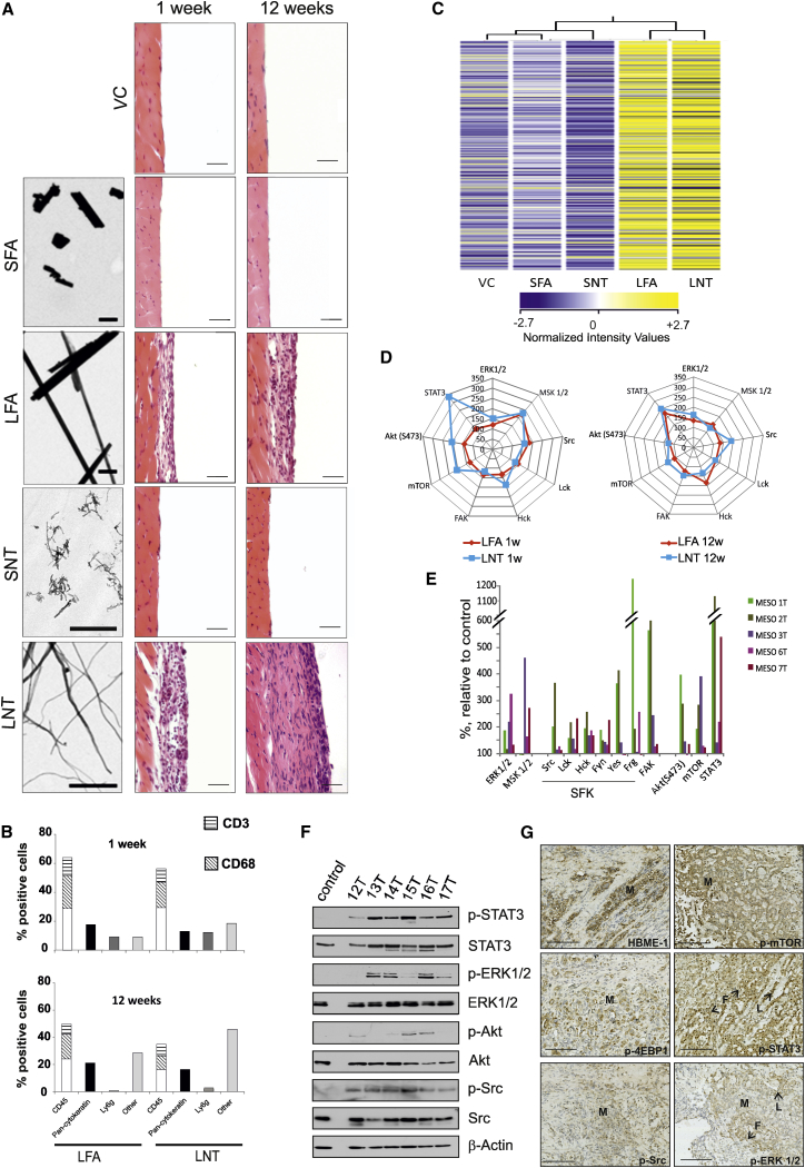
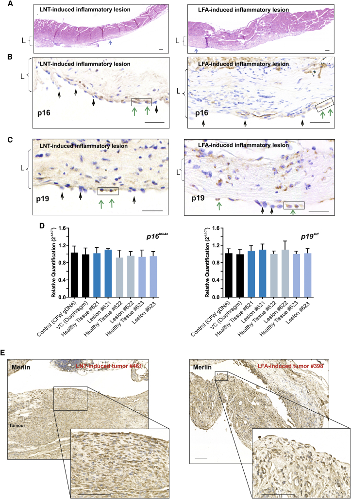
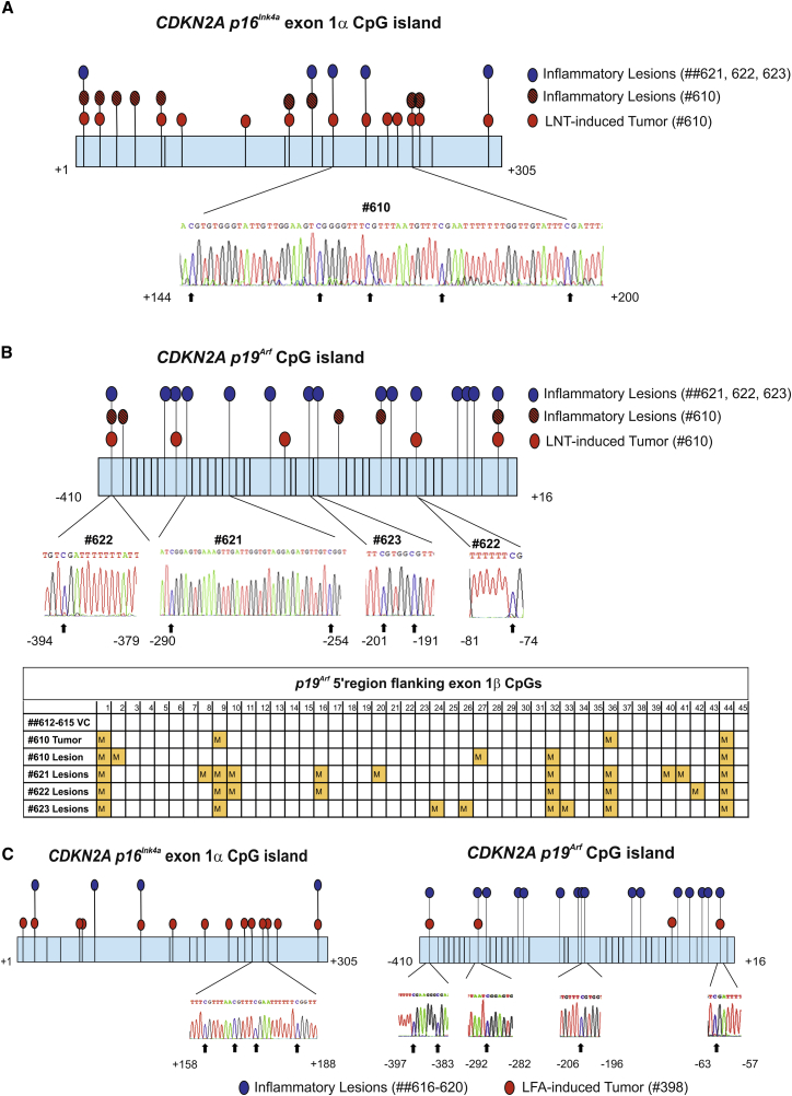
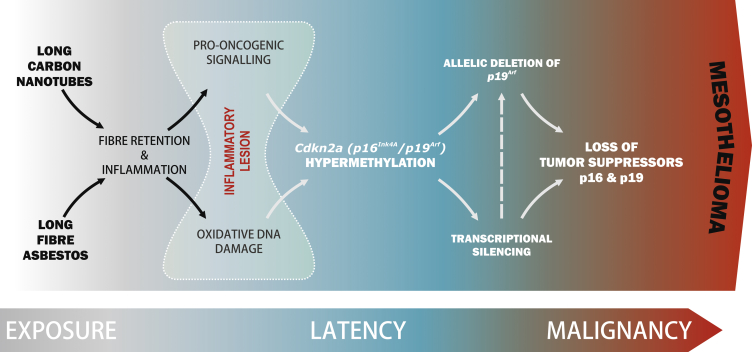
Mesothelioma is a fatal tumor of the pleura and is strongly associated with asbestos exposure. The molecular mechanisms underlying the long latency period of mesothelioma and driving carcinogenesis are unknown. Moreover, late diagnosis means that mesothelioma research is commonly focused on end-stage disease. Although disruption of the CDKN2A (INK4A/ARF) locus has been reported in end-stage disease, information is lacking on the status of this key tumor suppressor gene in pleural lesions preceding mesothelioma. Manufactured carbon nanotubes (CNTs) are similar to asbestos in terms of their fibrous shape and biopersistent properties and thus may pose an asbestos-like inhalation hazard. Here we show that instillation of either long CNTs or long asbestos fibers into the pleural cavity of mice induces mesothelioma that exhibits common key pro-oncogenic molecular events throughout the latency period of disease progression. Sustained activation of pro-oncogenic signaling pathways, increased proliferation, and oxidative DNA damage form a common molecular signature of long-CNT- and long-asbestos-fiber-induced pathology. We show that hypermethylation of p16/Ink4a and p19/Arf in CNT- and asbestos-induced inflammatory lesions precedes mesothelioma; this results in silencing of Cdkn2a (Ink4a/Arf) and loss of p16 and p19 protein, consistent with epigenetic alterations playing a gatekeeper role in cancer. In end-stage mesothelioma, silencing of p16/Ink4a is sustained and deletion of p19/Arf is detected, recapitulating human disease. This study addresses the long-standing question of which early molecular changes drive carcinogenesis during the long latency period of mesothelioma development and shows that CNT and asbestos pose a similar health hazard.

Keywords: CDKN2A; asbestos; carbon nanotubes; epigenetics; hypermethylation; mesothelioma; toxicity; tumor suppressor genes.


Figures

Figure 1
Figure 1
Asbestos and CNT Fiber-Induced Pleural Lesions Exhibit Common Length-Dependent Molecular Changes (A) Left: transmission electron microscopy (TEM) images of the fiber panel; scale bars, 1 μm. Right: hematoxylin and eosin (H&E) images of the chest wall of mice at 1 and 12 weeks post-injection of the fiber panel (SFA, SNT, LFA, or LNT) compared to VC. Scale bars, 20 μm. (B) Cell types within the lesion were quantified (700–1,000 cells per cell marker, per treatment group; n = 4 per group) according to immunostaining (Figure S2) at 1 week and 12 weeks. (C) Gene expression pattern in control and fiber-exposed animals at 12 weeks post-injection. The heatmap displays the expression level of mRNAs extracted from the diaphragms of animals exposed to SFA, SNT, LFA, or LNT and VC (n = 4 per group). Legend bar shows the color code for the normalized intensity values. (D and E) Antibody array-based kinome profiling. (D) Common pattern of kinase activation induced by exposure to LFA and LNT at 1 and 12 weeks relative to VC (100%). (E) Activation of signaling pathways in human mesothelioma tissue from five patients is shown relative to kinase activity in normal primary mesothelial cells (control). (F) Human mesothelioma tissue was analyzed for phospho-STAT3 (Y705), phospho-ERK1/2 (T202/204), phospho-Akt (S473), and phospho-Src (Y418) by western blotting and compared to normal primary mesothelial cells (control). Representative data from six patients are shown. (G) Immunostaining of signaling proteins in paraffin-embedded sections of human mesothelioma tissue in relation to cancerous (M) and non-cancerous (L, lymphocytes; F, fibroblasts) cells. Representative data from three patients are shown. Scale bars, 100 μm. See also Figures S1–S3 and Tables S1–S3.
Figure 2
Figure 2
Sustained Inflammation and Activation of Pro-oncogenic Signaling Pathways in LFA- and LNT-Induced Lesions at 6 Months Post-injection (A) Cell types in LFA- and LNT-induced inflammatory lesions at 6 months post-injection were quantified by immunostaining with a panel of cell markers (pan-cytokeratin, mesothelial cells; CD68, macrophages; CD45, leukocytes; CD3, T cells; Ly6g, granulocytes). (B) Specific areas of tissue were isolated from fresh frozen diaphragms (n = 4) by laser microdissection in order to examine gene expression levels in different cell types (muscle, mesothelium, and lesion). For validation of the cell-type selection, the expression level of the cell markers mesothelin and CD68 in the different areas sampled was measured by qPCR. The expression level of the genes encoding STAT3, IL-6, and PI3K was examined by qPCR in muscle, mesothelium, and lesion (where present) microdissected from mice exposed to VC, LFA, or LNT at 12 weeks post-injection. p < 0.05, ∗∗p < 0.01. (C) Activation of signaling pathways in the chest wall tissue of mice 6 months post-injection was analyzed by immunostaining. Positive staining for signaling proteins was observed in both mesothelial and non-mesothelial cells (black arrows). Scale bar, 20 μm. See also Figure S4.
Figure 3
Figure 3
Progressing LNT-Induced Inflammatory Lesions Display Increased Proliferation and DNA Damage (A) Representative H&E-stained sections of pleurae from VC-, LFA-, and LNT-exposed mice at 6 months post-injection. Callouts show plump proliferating mesothelial cells (M) on the pleural surface of LFA- and LNT-exposed mice (positive for pan-cytokeratin; proliferation marker, Ki-67; and mitotic marker, p-Histone H3). Scale bars, 50 μm. (B) Increased proliferation in lesions of LFA- and LNT-exposed mice at 12 weeks and 6 months post-injection compared to VC, quantified by cells stained positively for Ki-67 and p-Histone H3 (700–1,000 cells per cell marker, per animal; n = 3 per group). (C) Sustained DNA damage in LFA- and LNT-induced lesions. The percentage of genomic DNA containing 8-hydroxy-2′-deoxyguanosine (8-OHdG) progressively increased in diaphragms of mice exposed to LFA or LNT compared to VC (n = 4 per group). Graphs (B and C) show mean ± SD; p < 0.05, ∗∗p < 0.01 (two-tailed Student’s t test).
Figure 4
Figure 4
LNT- and LFA-Induced Inflammatory Lesions Progress to Malignant Mesothelioma with Disruption of Cdkn2a Gene and Encoded Proteins (A) LNT-induced mesothelioma at 12 months post-injection (animal ID: no. 610). The callout shows the mesothelioma composed of pleomorphic epithelioid tumor cells infiltrating into the underlying muscle (arrow). Adjacent to the tumor is an inflammatory lesion (L). Scale bars, 100 μm. (B) Immunostaining of LNT-induced mesothelioma (animal ID: no. 610). Tumor cells stained positively for the mesothelial cell markers pan-cytokeratin and WT1 (arrows); the tumor areas stained positively (arrows) or negatively (circle) for the Cdkn2a-encoded proteins p16 and p19. Scale bars, 100 μm. (C) Immunostaining of LNT-induced tumor (animal ID: no. 610) for p16 and p19. Negatively (circled) and positively (red arrows) stained areas were dissected and collected by power-assisted laser micro-dissection (PALM) for gDNA extraction and qPCR analysis. Callouts show a subsequent crystal violet-stained section of tumor after collection of selected areas. Scale bars, 100 μm. (D) Relative quantification (mean of 2−ΔΔCT) of p16Ink4a and p19Arf gene copy number in gDNA from micro-dissected tumor and healthy tissue from the same animal or VC, showing allelic loss of p19Arf in p19-negative tumor areas compared to controls or p19-positive tumor areas. Graphs show mean ± SD; p < 0.05 (significant difference is defined by Z score analysis; see also STAR Methods). (E) Immunostaining of LFA-induced tumor (animal ID: no. 398) for p16 and p19 protein. Callouts show positively stained areas (circled) in the upper panels and negatively stained areas in the lower panels that were dissected and collected by PALM for gDNA extraction and qPCR analysis (see Figure S6C). Scale bars, 100 μm. See also Figures S5–S7.
Figure 5
Figure 5
Chronic Inflammatory Lesions Induced by Long Fibers Display Loss of p16 and 19 Expression (A) Representative H&E-stained sections of diaphragm from LNT- and LFA-exposed animals that did not have tumors at the 1 year study endpoint, displaying extensive chronic inflammatory lesions (L) with fibrosis, infiltrating inflammatory cells, and plump reactive mesothelial cells on the surface of pleural lesions (blue arrows). Scale bars, 100 μm. (B) Loss of p16 expression in mesothelial cells in LNT- and LFA-induced inflammatory lesions. Representative images of immunostaining for p16 show predominantly negatively stained (black arrows) and only a few (boxed) positively stained (green arrows) mesothelial cells. Scale bars, 100 μm. (C) Loss of p19 protein expression in mesothelial cells in LNT- and LFA-induced inflammatory lesions. Representative images of immunostaining for p19 show predominantly negatively stained (black arrows) and only a few (boxed) positively stained (green arrows) mesothelial cells. Scale bars, 100 μm. (D) Relative quantification (mean of 2−ΔΔCT) of p16Ink4a and p19Arf gene copy number by qPCR analysis in gDNA isolated from positively and negatively stained mesothelial cells in the LNT-induced inflammatory lesions (animal IDs: nos. 621, 622, and 623) compared to controls. (E) Immunostaining for the NF2-encoded protein Merlin in LNT (animal ID: no. 461) and LFA (animal ID: no. 398) tumors. Representative images show positive cytoplasmic staining of tumor areas. Scale bars, 100 μm.
Figure 6
Figure 6
Chronic Inflammatory Lesions and Malignant Mesothelioma Induced by LFA and LNT Display Hypermethylation of the Cdkn2a (p16 Ink4a/p19Arf) Locus (A) Schematic representation of the hypermethylation profile of the p16Ink4a CpG island in exon 1α determined by bisulphite sequencing of gDNA. All CpG dinucleotides are shown by vertical lines. Hypermethylated CpG sites were identified in gDNA extracted from p16-negative areas of LNT-induced tumor (animal ID: no. 610; red-filled circles), in gDNA isolated from mesothelial cells in inflammatory non-neoplastic lesions from the same animal (hatched circles), and in gDNA from mesothelial cells in chronic inflammatory lesions of LNT-exposed animals that did not develop tumors at the 1 year study endpoint (animal IDs: nos. 621, 622, ad 623; blue-filled circles). The call-out shows a region of bisulphite converted sequence with unconverted cytosines (arrows). (B) Schematic representation of the hypermethylation status of the 5′ p19Arf CpG island located upstream of exon 1β determined by bisulphite sequencing. All CpG dinucleotide positions are shown by vertical lines. Hypermethylated CpG sites were identified in p19-negative areas of LNT-induced tumor (no. 610; red-filled circles), in mesothelial cells isolated from inflammatory non-neoplastic lesions of the same animal (hatched circles), and in mesothelial cells isolated from chronic inflammatory lesions of LNT-exposed animals that did not develop tumors at the 1 year study endpoint (animal IDs: nos. 621, 622, and 623; blue-filled circles). Callouts show examples of unconverted cytosines in hypermethylated CpGs. Results are summarized in the table, where each yellow-filled square represents multiple sequencing runs (unconverted CpG detected in 10–60 clones compared to none in VC). (C) Schematic representation of the hypermethylation profile of the p16Ink4a CpG island in exon 1α and the 5′ p19Arf CpG island located upstream of exon 1β determined by bisulphite sequencing. Hypermethylated CpG sites were identified in gDNA extracted from p16-negative areas of LFA-induced tumor (animal ID: no. 398; red-filled circles) and in gDNA from mesothelial cells in chronic inflammatory lesions of LFA-exposed animals that did not develop tumors at the 1 year study endpoint (animal IDs: nos. 616–620; blue-filled circles). The callout shows a region of bisulphite converted sequence with unconverted cytosines. See also Figure S7.
Figure 7
Figure 7
LNTs Replicate Asbestos-Induced Mesothelioma with Disruption of the Tumor Suppressor Gene Cdkn2a (Ink4a/Arf) Schematic depicting the sequence of events in the pleurae of animals exposed to LFA or LNT showing commonality in LFA- and LNT-induced disease progression that replicates mesothelioma development in humans.

Comment in

References

    1. Murphy F.A., Poland C.A., Duffin R., Al-Jamal K.T., Ali-Boucetta H., Nunes A., Byrne F., Prina-Mello A., Volkov Y., Li S. Length-dependent retention of carbon nanotubes in the pleural space of mice initiates sustained inflammation and progressive fibrosis on the parietal pleura. Am. J. Pathol. 2011;178:2587–2600. - PMC - PubMed
    1. Nagai H., Toyokuni S. Biopersistent fiber-induced inflammation and carcinogenesis: lessons learned from asbestos toward safety of fibrous nanomaterials. Arch. Biochem. Biophys. 2010;502:1–7. - PubMed
    1. Lecomte C., Andujar P., Renier A., Kheuang L., Abramowski V., Mellottee L., Fleury-Feith J., Zucman-Rossi J., Giovannini M., Jaurand M.C. Similar tumor suppressor gene alteration profiles in asbestos-induced murine and human mesothelioma. Cell Cycle. 2005;4:1862–1869. - PubMed
    1. Guo G., Chmielecki J., Goparaju C., Heguy A., Dolgalev I., Carbone M., Seepo S., Meyerson M., Pass H.I. Whole-exome sequencing reveals frequent genetic alterations in BAP1, NF2, CDKN2A, and CUL1 in malignant pleural mesothelioma. Cancer Res. 2015;75:264–269. - PubMed
    1. Lo Iacono M., Monica V., Righi L., Grosso F., Libener R., Vatrano S., Bironzo P., Novello S., Musmeci L., Volante M. Targeted next-generation sequencing of cancer genes in advanced stage malignant pleural mesothelioma: a retrospective study. J. Thorac. Oncol. 2015;10:492–499. - PubMed

MeSH terms