. 2017 Dec:60:89-101.
doi: 10.1016/j.dnarep.2017.10.009. Epub 2017 Oct 24.

Natural product β-thujaplicin inhibits homologous recombination repair and sensitizes cancer cells to radiation therapy

Affiliations

Natural product β-thujaplicin inhibits homologous recombination repair and sensitizes cancer cells to radiation therapy

Lihong Zhang et al. DNA Repair (Amst). 2017 Dec.

Abstract

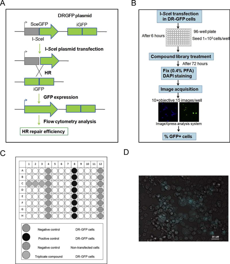
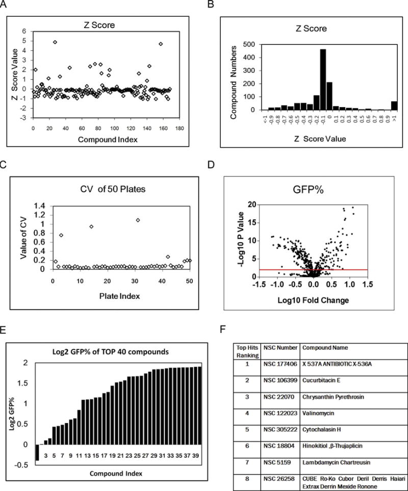
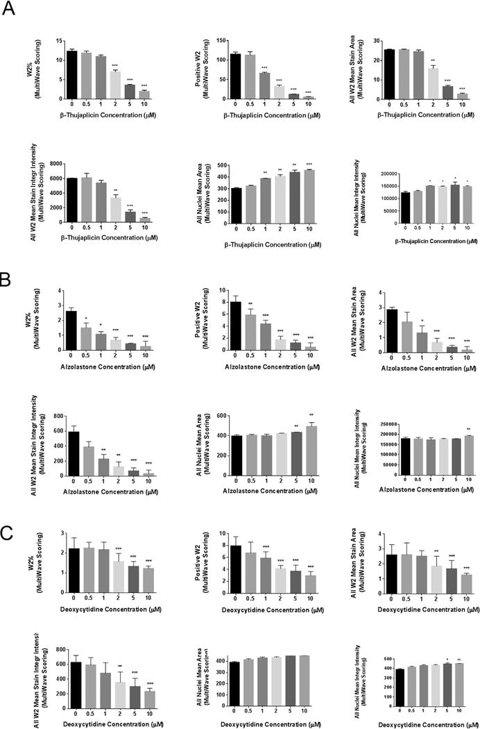
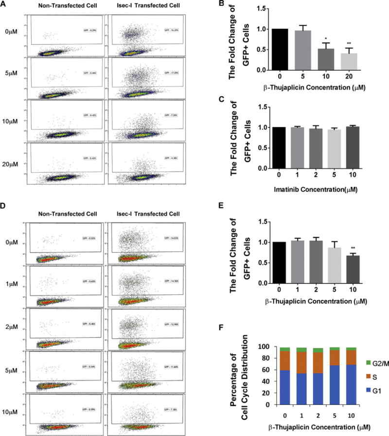
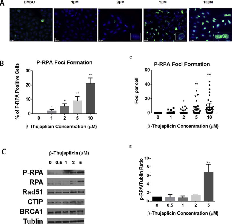
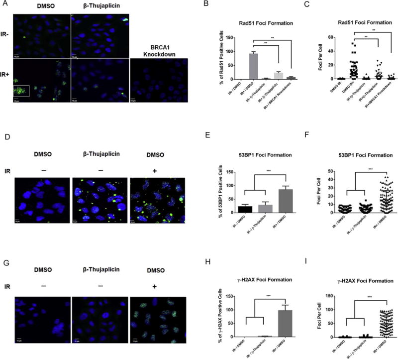
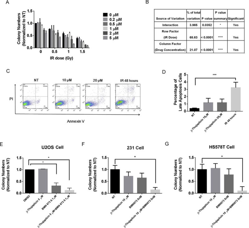
Investigation of natural products is an attractive strategy to identify novel compounds for cancer prevention and treatment. Numerous studies have shown the efficacy and safety of natural products, and they have been widely used as alternative treatments for a wide range of illnesses, including cancers. However, it remains unknown whether natural products affect homologous recombination (HR)-mediated DNA repair and whether these compounds can be used as sensitizers with minimal toxicity to improve patients' responses to radiation therapy, a mainstay of treatment for many human cancers. In this study, in order to systematically identify natural products with an inhibitory effect on HR repair, we developed a high-throughput image-based HR repair screening assay and screened a chemical library containing natural products. Among the most interesting of the candidate compounds identified from the screen was β-thujaplicin, a bioactive compound isolated from the heart wood of plants in the Cupressaceae family, can significantly inhibit HR repair. We further demonstrated that β-thujaplicin inhibits HR repair by reducing the recruitment of a key HR repair protein, Rad51, to DNA double-strand breaks. More importantly, our results showed that β-thujaplicin can radiosensitize cancer cells. Additionally, β-thujaplicin sensitizes cancer cells to PARP inhibitor in different cancer cell lines. Collectively, our findings for the first time identify natural compound β-thujaplicin, which has a good biosafety profile, as a novel HR repair inhibitor with great potential to be translated into clinical applications as a sensitizer to DNA-damage-inducing treatment such as radiation and PARP inhibitor. In addition, our study provides proof of the principle that our robust high-throughput functional HR repair assay can be used for a large-scale screening system to identify novel natural products that regulate DNA repair and cellular responses to DNA damage-inducing treatments such as radiation therapy.

Keywords: DNA repair; Homologous recombination; PARP inhibitor; Radiosensitizer; β-thujaplicin.


Conflict of interest statement

Disclosure of conflict of interest

The authors declare that there are no conflicts of interest.

Figures

Fig. 1
Fig. 1
Designation of high-throughput image-based screening assay. A. Schematic diagram of DR-GFP cell-based functional HR repair assay. B. Schematic representation of the high-throughput screening. C. Plate layout designation. D. Automated imaging of U2OS cells engineered to permit assessment of the efficiency of HR repair. Cells expressing green fluorescent protein (GFP) indicating successful HR repair.
Fig. 2
Fig. 2
Performace of high-throughput image-based screening assay. A. Z score scatter plot of screening in NCI Natural Compounds Set III plates. B. Z score distribution of screening (NCI Natural Compounds Set III and Mechanistic Diversity Set II plates). C. CV scatter plot of 50 plates in the screening (NCI Natural Compounds Set III and Mechanistic Diversity Set II plates). D. Volcano plot of screening results (NCI Natural Compounds Set III and Mechanistic Diversity Set II plates). E. Top 40 hits waterfall plot in the primary screening (NCI Natural Compounds Set III and Mechanistic Diversity Set II plates). F. The compound list of top 8 positive hits in NCI Natural Compounds Set III plates.
Fig. 3
Fig. 3
HR repair inhibitor hits validation in a secondary screening assay. A. Secondary screening validation of positive hit β-thujaplicin. B. Secondary screening validation of positive hit Alzolastone. C. Secondary screening validation of positive hit Deoxycytidine. The multiwave scoring of W2 percentage, the number of W2 positve cells, the W2 mean stain area, and the W2 mean stain integrity intensity decreased significantly with increasing hits’ concentration(0, 0.5, 1, 2, 5, 10 μM). (W2 indicating wavelength 2 using by the analysis system to detect GFP signal by microscopy). All nuclei mean area and all nuclei mean integrity intensity increased significantly with increasing hits’ concentration. Error bars represent SD.*P < 0.05, **P < 0.01, ***P < 0.001.
Fig. 4
Fig. 4
Final confirmation of β-thujaplicin’s HR repair inhibition effect by flow cytometry. A. β-thujaplicin decreases HR repair efficiency. Representative flow cytometry profiles show a significant decrease of GFP-positive cells after treatment with different doses of β-thujaplicinas (0, 5, 10, 20 μM) indicated by I-SceI HR reporter assay. B. Quantitation of β-thujaplicin’s inhibitory effect on HR. β-thujaplicin decreases the fold change of GFP positive cells significantly at the concentration of 10 and 20 μM. Error bars represent SD. *P < 0.05, **P < 0.01. C. Quantitation of negative control imatinib’s effect on HR. Imatinib doesn’t change the fold change of GFP positive cells at different doses. Error bars represent SD. *P < 0.05, **P < 0.01. D. In contact inhibition assay (add β-thujaplicin or DMSO when cells reach 90% confluency), β-thujaplicin decreases the fold change of GFP positive cells. Representative flow cytometry profiles show a significant decrease of the fold change of GFP positive cells after treatment with different doses of β-thujaplicinas (0, 1, 2, 5, 10 μM) indicated by I-SceI HR reporter assay. E. Quantitation of β-thujaplicin’s inhibitory effect on HR. β-thujaplicin decreases the fold change of GFP positive cells significantly at the concentration of 10 μM. Error bars represent SD. *P < 0.05, **P < 0.01. F. In contact inhibition assay(add β-thujaplicinor DMSO when cells reach 90% confluency),cell cycle analysis shows that β-thujaplicin slightly increase the G1 cell cycle distribution at the concentration of 5 μM and 10 μM, but doesn’t cause further increased G1 cell cycle arrest.
Fig. 5
Fig. 5
β-thujaplicin induces phosphorylation of RPA. A. Immunofluorescence staining of p-RPA32 foci (green) in U2OS cells 48 hours following treatment with DMSO or β-thujaplicin (1, 2, 5, or 10 μM). The nucleus is counterstained with DAPI (blue). B. Quantitation of percentage of p-RPA32 foci-positive cells shows β-thujaplicin significantly increases p-RPA32 foci formation at the concentration of 1, 2, 5, or 10 μM. Error bars represent SD from replicates. *P < 0.05, **P < 0.01. C. Quantitation of p-RPA32 foci numbers per cell shows β-thujaplicin significantly increases p-RPA32 foci formation per cell at the concentration of 2, 5, or 10 μM. Error bars represent SD from replicates. *P < 0.05, **P < 0.01, ***P < 0.001. D. Western blot analysis (p-RPA,RPA,Rad51,CTIP,BRCA1,Tublin) of U2OS cells pretreated with DMSO or β-thujaplicin at the indicated concentrations (0.5, 1, 2, 5 μM) for 72 h. E. Quantitation of P-RPA protein level shows β-thujaplicin significantly increases the P-RPA protein level at the concentration of 5 μM. Error bars represent SD from replicates (*P < 0.05, **P < 0.01).
Fig. 6
Fig. 6
A–C β-thujaplicin inhibits radiation-induced Rad51 foci formation. A. Immunofluorescence staining of Rad51 foci (green) in U2OS cells 8 h following treatment with β-thujaplicin (10 μM) followed 16 h later by exposure to IR (10 Gy). The nucleus is counterstained with DAPI (blue). B. Quantitation of percentage of Rad51 foci-positive cells shows β-thujaplicin significantly decreases Rad51 foci formation in U2OS cells following treatment with β-thujaplicin (10 μM) compared to DMSO followed 16 h later by exposure to IR (10 Gy). Error bars represent SD from replicates. *P < 0.05, **P < 0.01. C. Quantitation of Rad51 foci numbers per cell shows β-thujaplicin significantly decreases Rad51 foci formation in U2OS cells following treatment with β-thujaplicin (10 μM) compared to DMSO followed 16 h later by exposure to IR (10 Gy). Error bars represent SD from replicates. *P < 0.05, **P < 0.01. D–I. β-thujaplicin does not cause accumulation of DSBs. D. Immunofluorescence staining of 53BP1 foci (green) in U2OS cells 2 h following treatment with β-thujaplicin (5 μM) or DMSO. The nucleus is counterstained with DAPI (blue). Positive control cells were exposed to IR (10 Gy). E. Quantitation of percentage of 53BP1 foci-positive cells shows β-thujaplicin doesn’t increases 53BP1 foci formation in U2OS cells following treatment with β-thujaplicin (5 μM) compared to DMSO. Error bars represent SD from replicates.*P < 0.05, **P < 0.01, ***P < 0.001. F. Quantitation of 53BP1 foci numbers per cell shows β-thujaplicin doesn’t increase 53BP1 foci formation in U2OS cells following treatment with β-thujaplicin (5 μM) compared to DMSO. Error bars represent SD from replicates.*P < 0.05, **P < 0.01, ***P < 0.001. G. Immunofluorescence staining of γ-H2AX foci (green) in U2OS cells 2 h following treatment with β-thujaplicin (5 μM) or DMSO. The nucleus is counterstained with DAPI (blue). Positive control cells were exposed to IR (10 Gy). H. Quantitation of percentage of γ-H2AX foci-positive cells shows β-thujaplicin doesn’t increase γ-H2AX foci formation in U2OS cells following treatment with β-thujaplicin (5 μM) compared to DMSO. Error bars represent SD from replicates. *P < 0.05, **P < 0.01, ***P < 0.001. I. Quantitation of γ-H2AX foci numbers per cell shows β-thujaplicin doesn’t increase γ-H2AX foci formation in U2OS cells following treatment with β-thujaplicin (5 μM) compared to DMSO. Error bars represent SD from replicates. *P < 0.05, **P < 0.01, ***P < 0.001.
Fig. 7
Fig. 7
Effect of β-thujaplicin on radiosensitivity or combination of PARP inhibitor. A. Survival of U2OS cells treated with DMSO or β-thujaplicin at the indicated concentration (0.2, 0.5, 1, 2, 5 μM) and either not exposed to IR or exposed to IR at the indicated doses (0.5, 1, 1.5 Gy). B. Statistical analysis of impact of interaction between β-thujaplicin concentration and IR dose on cell survival. *P < 0.05, ****P < 0.0001. C. The apoptotic profile of U2OS cells treated with DMSO or β-thujaplicin at the indicated doses for 24 h. Positive control cells were exposed to IR (10 Gy). D. The quantitative data for apoptosis assay from three independent experiments shows β-thujaplicin at the indicated concentration (10, 20 μM) doesn’t induce significant cell apoptosis. ***P < 0.001. E. Survival assay of U2OS cells treated with DMSO, β-thujaplicin (5 μM), BMN673 (0.1 μM), and the combination group. F. Survival assay of breast cancer MDA-MB-231 cells treated with DMSO,β-thujaplicin (10 μM), BMN673 (5 nM),and the combination group. G. Survival of breast cancer HS578T cells treated with DMSO, β-thujaplicin (10 μM), BMN673 (5 nM), and the combination group.

Similar articles

Cited by

References

    1. Ward E, DeSantis C, Robbins A, Kohler B, Jemal A. Childhood and adolescent cancer statistics 2014. CA: Cancer J Clin. 2014;64:83–103. - PubMed
    1. Feng H, Wang J, Chen W, Shan B, Guo Y, Xu J, Wang L, Guo P, Zhang Y. Hypoxia-induced autophagy as an additional mechanism in human osteosarcoma radioresistance. J Bone Oncol. 2016;5:67–73. - PMC - PubMed
    1. Schwarz R, Bruland O, Cassoni A, Schomberg P, Bielack S. The role of radiotherapy in oseosarcoma. Cancer Treat Res. 2009;152:147–164. - PubMed
    1. Luetke A, Meyers PA, Lewis I, Juergens H. Osteosarcoma treatment - where do we stand? A state of the art review. Cancer Treatment Rev. 2014;40:523–532. - PubMed
    1. Bielack SS, Hecker-Nolting S, Blattmann C, Kager L. Advances in the management of osteosarcoma. F1000Research. 2016;5:2767. - PMC - PubMed

Publication types

MeSH terms