. 2017 Nov 7;12(11):e0187472.
doi: 10.1371/journal.pone.0187472. eCollection 2017.

Adjustment of the dynamic weight distribution as a sensitive parameter for diagnosis of postural alteration in a rodent model of vestibular deficit

Affiliations

Adjustment of the dynamic weight distribution as a sensitive parameter for diagnosis of postural alteration in a rodent model of vestibular deficit

Brahim Tighilet et al. PLoS One. .

Abstract

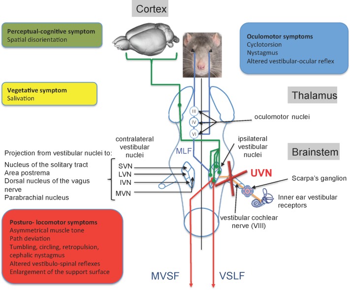
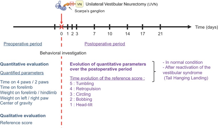
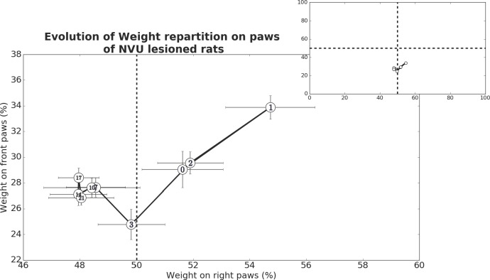
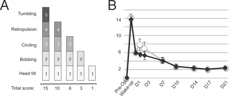
Vestibular disorders, by inducing significant posturo-locomotor and cognitive disorders, can significantly impair the most basic tasks of everyday life. Their precise diagnosis is essential to implement appropriate therapeutic countermeasures. Monitoring their evolution is also very important to validate or, on the contrary, to adapt the undertaken therapeutic actions. To date, the diagnosis methods of posturo-locomotor impairments are restricted to examinations that most often lack sensitivity and precision. In the present work we studied the alterations of the dynamic weight distribution in a rodent model of sudden and complete unilateral vestibular loss. We used a system of force sensors connected to a data analysis system to quantify in real time and in an automated way the weight bearing of the animal on the ground. We show here that sudden, unilateral, complete and permanent loss of the vestibular inputs causes a severe alteration of the dynamic ground weight distribution of vestibulo lesioned rodents. Characteristics of alterations in the dynamic weight distribution vary over time and follow the sequence of appearance and disappearance of the various symptoms that compose the vestibular syndrome. This study reveals for the first time that dynamic weight bearing is a very sensitive parameter for evaluating posturo-locomotor function impairment. Associated with more classical vestibular examinations, this paradigm can considerably enrich the methods for assessing and monitoring vestibular disorders. Systematic application of this type of evaluation to the dizzy or unstable patient could improve the detection of vestibular deficits and allow predicting better their impact on posture and walk. Thus it could also allow a better follow-up of the therapeutic approaches for rehabilitating gait and balance.


Conflict of interest statement

Competing Interests: The authors have declared that no competing interests exist.

Figures

Fig 1
Fig 1. Anatomo-functional organization of the central vestibular system and functional consequences of the unilateral vestibular neurectomy.
The vestibular nerve contacts the sensory cells within the vestibular endorgans (semicircular canals, utricle and saccule) and projects ipsilaterally on four vestibular nuclei (VN): the medial (MVN), inferior VN (IVN), lateral (LVN) and superior (SVN) vestibular nuclei. The VNs are located in the dorso-lateral part of the bulbo-protuberantial junction of the brainstem under the floor of the IVth ventricle. They form the first relay of the vestibular-ocular, vestibulospinal and vestibulo-vegetative reflexes and the vestibule-cortical ascending pathways involved in spatial orientation. VNs are the origin of pre-motor messages (control of the ocular and somatic musculature) involved in the regulation of the posture and the stabilization of the gaze during head movements. They project, through the medial longitudinal fasciculus (MLF) on oculomotor nuclei (oculomotor nucleus III, cochlear nucleus or abducens IV) whose motoneurons control the eye muscles to produce compensatory reactions of the eye for stabilizing the images on the retina during head movements. Vestibulo-spinal projections, originating from the ipsilateral LVN, reach all medullary stages via the lateral vestibulospinal fasciculus (VSLF). Contralateral projections median of the median, inferior and lateral VN constitute the median vestibulospinal fasciculus (MVSF) and control the motoneurons of the neck and the upper part of the body axis. VNs receive cerebellar, medullary (proprioceptive) and visual (optokinetic) afferents from contralateral vestibular nuclei and cortical areas. In Human, several cortical areas are involved in the reception of multisensorial vestibular messages (3a area 2v area, parietal-insular vestibular cortex) running through the thalamo-cortical fasciculus. In rodents, vestibular input to the cortex is widespread, affecting many functionally different areas. Some of these areas are homologous with primate vestibular maps (e.g., cingulate and somatosensory cortex), whereas others seem to be specific to rodents (e.g., medial prefrontal cortex). Given this anatomical-functional organization of the vestibular system and its projection targets, unilateral vestibular nerve section induces a quadruple syndrome: posture-locomotor (blue), oculomotor (green), vegetative (pink) and perceptive-cognitive (red). Adapted from [6].
Fig 2
Fig 2. Illustration of the dynamic weigh bearing device used to monitor the dynamic weight distribution in rodents.
A: high definition camera (a), coverlid and camera support (b), assembled coverlid (c), force sensors and glass cage (d), assembled system (e). B: Dynamic weigh bearing system in acquisition condition. The rat is free moving in the cage. Its contact points with the captors and the video of its displacement are sent on line to the acquisition software.
Fig 3
Fig 3. Illustration of the monitoring set up in condition of acquisition.
A: a colour is assigned to each area of contact between the animal body and the force sensors. The coloured bar below represents the acquisition time (here 5min). The green corresponds to the time analysed, the red to the non-analysable segments. The grey attachment is a visual marker (representing the electrical connection with the interface) that allows juxtaposition of both the digitized false colour image and the picture of the rat. The grey bar is the cursor of the analysed image. B: Picture taken from the video tracking of the analysed animal.
Fig 4
Fig 4. Details of the procedure used to evaluate the alterations of both the posturo locomotor behaviour and the dynamic weigh distribution during the pre-operative and postoperative periods.
Fig 5
Fig 5. Representation of the time spent on two and four paws in control rats and after 1 and 10 days after UVN.
Left: control rats. Middle: 1 day after UVN. This situation corresponds to the acute stage of the vestibular syndrome. Right: 10 days after UVN. This situation corresponds to the compensated stage of the vestibular syndrome. Pictures below illustrate the righting position that characterizes the exploration behaviour (right and left) and the body position characteristic of the acute vestibular syndrome (Middle). At this stage, the rat favours a support surface on the intact side probably due to the increase of the muscular tonus of the paws on the intact side and the simultaneous loss of tonicity on the injured side.
Fig 6
Fig 6. Representation of the time spent on two and four paws for the rats in normal situation and after the UVN.
The time spent on two and four paws is represented as a percentage of time compared to the time analysed over the 5 minutes of acquisition. In control situation (A) there is a statistically significant increase in the time spent on four paws and a significant decrease in the time spent on two paws over the first three days after the lesion, relative to the pre-operative condition (a; p <0.05). These changes are followed by a return to preoperative values from D7 in both cases. In reactivated condition (B), there is a statistically significant increase in the time spent on four paws and a significant decrease in the time spent on two paws over the first three days after the lesion, relative to the pre-operative condition (a; p <0.05). Statistically significant change relative to the pre-operative condition is prolonged over the D7-D21 period, only for the time spent on two paws. Over this period, drastic reduction of visual and proprioception inputs induces significant changes (b; p <0.05) in both the time spent on two and four paws relative to the control situation.
Fig 7
Fig 7. Representation of the time spent on the front right and left paws before and after the UVN.
A: In control situation, statistically significant increase in the time spent on the front right and left paws is observed over the first three days after the lesion, relative to the pre-operative condition (a; p<0.05). Over the D7-D21 period, only the time spent on the front left paws remains significantly different relative to the control situation (c; p<0,05). In reactivated condition (B), statistically significant change relative to the pre-operative condition is prolonged over the D7-D21 period, only for the time spent on front left paw. Over this period, statistically significant change (b; p<0.05) in both the time spent on the right and left paws relative to the control situation is observed.
Fig 8
Fig 8. Representation of the weight distribution on each leg before and after UVN.
A-B: Antero-posterior axis. Normal situation (A), after reactivation of the vestibular syndrome (B). The rat weight is transferred forward over the first few days, with a statistically significant increase at P1 in the weight applied to the front paws and a simultaneous decrease of the weight on the hind paws relative to pre-operative condition (a; p<0,05). Reactivation of the vestibular syndrome does not significantly affect the antero-posterior weight distribution. C-D: Lateral axis. Normal situation (C), after reactivation (D). The rats apply more weight on the contralateral (right) side to the UVN the first two days, with a tendency for simultaneously reducing the weight on the opposite paw. This trend is reversed from P7 and maintained over the 3 weeks period monitored (in both cases: statistically significant difference relative to the pre-operative condition a; p<0,05). Reactivation of the syndrome affects only the weight distribution until D2 (c, p<0,05).
Fig 9
Fig 9. Representation of the barycenter evolution over time following UVN.
The abscissa represents the weight percentage on the right paws (cumulated weight on front and rear right paws in percent of the cumulated weight on the four paws), while the ordinate represents the animal weight percentage on the front paws (cumulated weight on front left and front right paws in percent of the cumulated weight on the four paws). The top right panel represents the same dataset than the larger down panel, it only shows the full axis of the figure, and pictures the short range in which our barycentre-like computation varies highlighting the recording of small postural disruptions with the DWB device. At D1, the animal weight shifted on the paws contralateral to the lesion (+3.11% on the right paws of the animal) and on the front paws (+4.83%). Each barycenter-like computation (white numbered dots) are mean barycenter-like calculated from 9 animals and taken from one recording session for each animal.
Fig 10
Fig 10. Qualitative assessment of the posturo-locomotor component of the vestibular syndrome.
(A) Illustration of the evaluation grid used in present study. A first state in which all symptoms are expressed is rated 15. A second stage in which the tumbling has gone is rated 10. A third state in which both the tumbling and the retropulsion behaviours are absent is rated 6. Then, two states (rated 3 and 1 respectively) related to states in which both bobbing and head tilt, or head tilt alone remained. (B) Illustration of the time course of the behavioural evaluation score involving various posturo-locomotor components of the vestibular syndrome according to the procedure described in Materials and Methods. The expression kinetics of the vestibular syndrome displays different phases: an acute phase of high intensity in the hours following the vestibular lesion (awakening and 4h), an intermediate phase of drastic reduction of the syndrome during the first post-lesion week (D1-D7), and a compensated phase (1–3 weeks), leading to almost complete disappearance of the symptoms. Each data point represents mean vestibular score, with error bars representing sem. Statistically significant difference on the behavioural score is only obtained at D1 and D3. Control (black line and square) and reactivated (white dots and grey line) conditions.

References

    1. Asher DL. Vestibular anatomy and physiology In: Northern JL. editor. Hearing Disorders. 2 Boston, MA: Little, Brown and Company; 1984; 195–204.
    1. Luxon LM. The anatomy and physiology of the vestibular system In: Dix MR, Hood JD editors. Vertigo. Chichester, UK: John Wiley & Sons, Ltd; 1984; 1–33.
    1. Jones SM, Jones TA, Mills KN, Gaines GC. Anatomical and physiological considerations in vestibular dysfunction and compensation. Semin Hear. 2009; 30(4): 231–241. doi: 10.1055/s-0029-1241124 - DOI - PMC - PubMed
    1. Angelaki DE, Cullen KE. Vestibular system: the many facets of a multimodal sense. Annu Rev Neurosci. 2008; 31: 125–150. doi: 10.1146/annurev.neuro.31.060407.125555 - DOI - PubMed
    1. Lacour M, Tighilet B. Plastic events in the vestibular nuclei during vestibular compensation: the brain orchestration of a deafferentation code. Rest Neurol Neurosci. 2010; 27: 1–17. - PubMed