. 2017 Nov 5;22(11):1904.
doi: 10.3390/molecules22111904.

The Oligomeric Form of the Escherichia coli Dps Protein Depends on the Availability of Iron Ions

Affiliations

The Oligomeric Form of the Escherichia coli Dps Protein Depends on the Availability of Iron Ions

Sergey Antipov et al. Molecules. .

Abstract

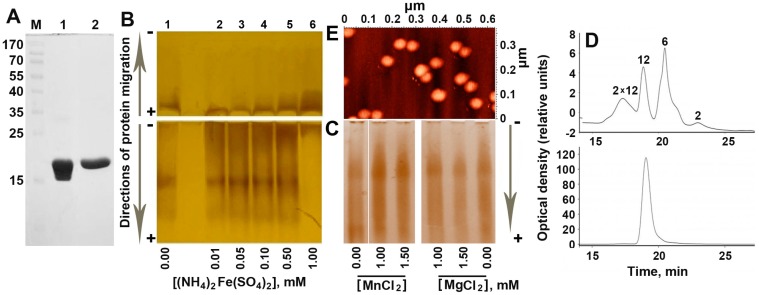
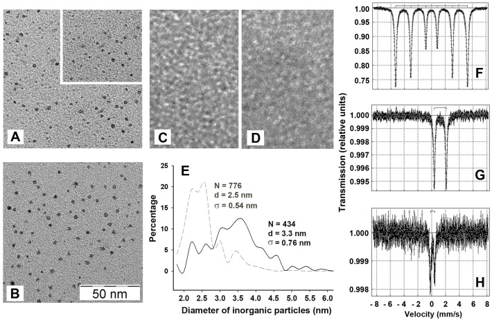
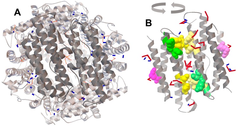
The Dps protein of Escherichia coli, which combines ferroxidase activity and the ability to bind DNA, is effectively used by bacteria to protect their genomes from damage. Both activities depend on the integrity of this multi-subunit protein, which has an inner cavity for iron oxides; however, the diversity of its oligomeric forms has only been studied fragmentarily. Here, we show that iron ions stabilize the dodecameric form of Dps. This was found by electrophoretic fractionation and size exclusion chromatography, which revealed several oligomers in highly purified protein samples and demonstrated their conversion to dodecamers in the presence of 1 mM Mohr's salt. The transmission electron microscopy data contradicted the assumption that the stabilizing effect is given by the optimal core size formed in the inner cavity of Dps. The charge state of iron ions was evaluated using Mössbauer spectroscopy, which showed the presence of Fe₃O₄, rather than the expected Fe₂O₃, in the sample. Assuming that Fe2+ can form additional inter-subunit contacts, we modeled the interaction of FeO and Fe₂O₃ with Dps, but the binding sites with putative functionality were predicted only for Fe₂O₃. The question of how the dodecameric form can be stabilized by ferric oxides is discussed.

Keywords: Dps proteins; Escherichia coli; Mössbauer spectroscopy; ferroxidase; molecular docking; oligomerization; transmission electron microscopy.


Conflict of interest statement

The authors declare no conflict of interest.

Figures

Figure 1
Figure 1
Oligomeric composition of the purified protein Dps. (A) The purity level of the protein obtained after ion exchange chromatography (lane 1) and gel filtration (lane 2) as revealed by SDS electrophoresis in 7.5% polyacrylamide gel. Protein marker lengths (M) are indicated in kDa. (B) Titration of purified Dps (100 μM calculated for monomers or <8.3 μM for dodecamers) with Mohr’s salt at pH 7.0. Lane 1—native Dps; lanes 2–6—fractional composition of the protein in the presence of 0.01–1 mM of Mohr’s salt, respectively. Vertical arrows on the left indicate the direction of electrophoretic fractionation. (C) The same for Dps titrated with MnCl2 or MgCl2. (D) Elution profiles of the Dps from the gel-filtration column in the HPLC system for the purified protein (top panel) and Dps loaded with 1 mM Mohr’s salt (bottom panel). The concentration of Dps used in this experiment was 1 mg/mL (53 μM for monomers). Oligomeric forms corresponding to the observed migration rate are indicated above the peaks. (E) Atomic force microscopy (AFM) image demonstrating aggregated Dps particles in the native protein. Since the freshly prepared protein samples required for AFM were fairly homogenous in oligomeric form [18], this method was not used to evaluate effects of Fe(NH4)2(SO4)2.
Figure 2
Figure 2
Characteristics of iron content in the inner cavity of Dps. (A) and (B) TEM images of inorganic particles taken from the native protein (1.64 μM with 126 Fe-ions per dodecamer) and Dps, loaded with 1 mM of Fe(NH4)2(SO4)2, respectively. Particles accounted for image analysis are countered by Altami Studio software (http://altamisoft.ru/products/altami_studio/). An insert in panel A shows an unprocessed image identical to the bottom part of the same panel. (C) apo-form of the Dps, prepared as described in Materials and Methods. (D) Image of 1 mM of Fe(NH4)2(SO4)2 alone. Scaling is identical for all images and is shown in (B). (E) Densitograms showing the size distribution of inorganic particles calculated with a resolution step of 0.2 nm. Dashed and solid lines correspond to native and iron-loaded proteins, respectively. N denotes the number of analyzed particles, d—average diameter of cores approximated by circles, σ—StD of d. (FH) Mössbauer spectra obtained for αFe, 1 mg of dry Fe(NH4)2(SO4)2 and for the Dps protein incubated with 1 mM Fe(NH4)2(SO4)2.
Figure 3
Figure 3
The positioning of FeO (blue rectangles) and Fe2O3 (red bent models) shown on the surface of dodecamer (A) and dimer (B). Accession code of the 3D model in the PDB: 1 dps [5]. Locations of Fe2O3 are accented in (B). The docking was performed independently for 50 molecules of FeO and Fe2O3, and two patterns of positional coordinates were superimposed on the 3D structural model of Dps (left panel). To show the distribution of iron oxides near the ferroxidase centers, two adjacent subunits (dark gray) were rotated for 180° (right panel). Yellow and green spheres show amino acids of ferroxidase and nucleation centers, respectively. Important for the integrity of Dps, R133 [30] is indicated in violet. The 3D structure of the Dps protein was visualized by RasMol 2.7.5 [31].

References

    1. Almiron M.A., Link J., Furlong D., Kolter R. A novel DNA-binding protein with regulatory and protective roles in starved Escherichia coli. Genes Dev. 1992;6:2646–2654. doi: 10.1101/gad.6.12b.2646. - DOI - PubMed
    1. Azam T.A., Ishihama A. Twelve species of the nucleoid-associated protein from Escherichia coli. Sequence recognition specificity and DNA binding affinity. J. Biol. Chem. 1999;274:33105–33113. doi: 10.1074/jbc.274.46.33105. - DOI - PubMed
    1. Talukder A.A., Ishihama A. Dps is a Stationary Phase-Specific Protein of Escherichia coli Nucleoid. Adv. Microbiol. 2014;4:1095–1104. doi: 10.4236/aim.2014.415120. - DOI
    1. Ishihama A., Kori A., Koshio E., Yamada K., Maeda H., Shimada T., Makinoshima H., Iwata A., Fujita N. Intracellular concentrations of 65 species of transcription factors with known regulatory functions in Escherichia coli. J. Bact. 2014;196:2718–2727. doi: 10.1128/JB.01579-14. - DOI - PMC - PubMed
    1. Grant R.A., Filman D.J., Finkel S.E., Kolter R., Hogle J.M. The crystal structure of Dps, a ferritin homolog that binds and protects DNA. Nat. Struct. Biol. 1998;5:294–303. doi: 10.1038/nsb0498-294. - DOI - PubMed

MeSH terms

LinkOut - more resources