. 2017 Sep;26(9):1505-1519.
doi: 10.1177/0963689717721221.

Adipose-Derived Mesenchymal Stem Cells from the Elderly Exhibit Decreased Migration and Differentiation Abilities with Senescent Properties

Affiliations

Adipose-Derived Mesenchymal Stem Cells from the Elderly Exhibit Decreased Migration and Differentiation Abilities with Senescent Properties

Meichen Liu et al. Cell Transplant. 2017 Sep.

Abstract

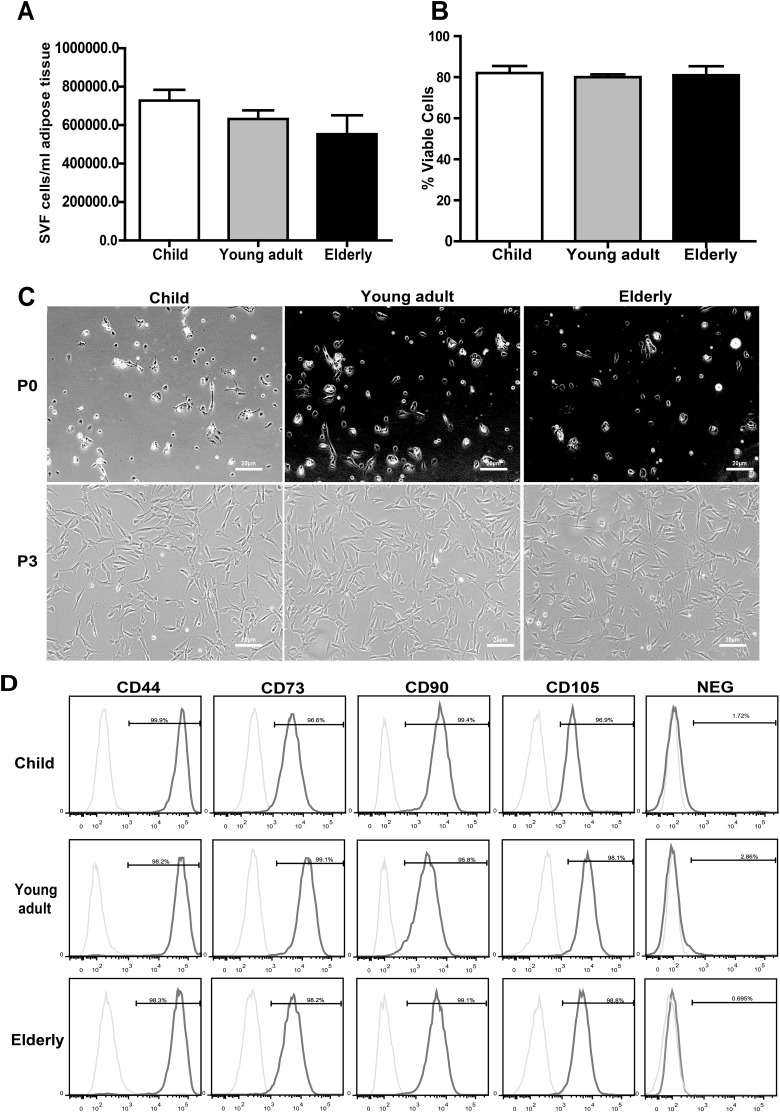
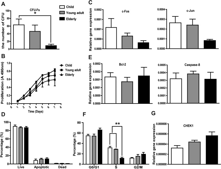
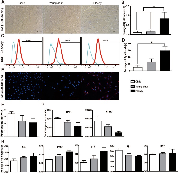
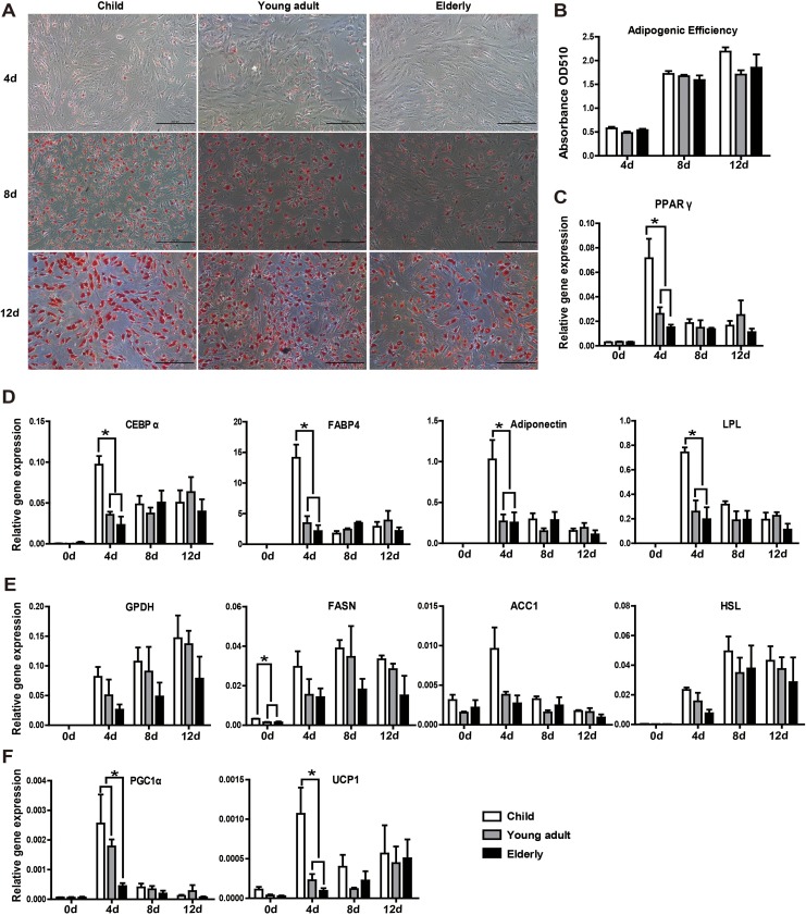
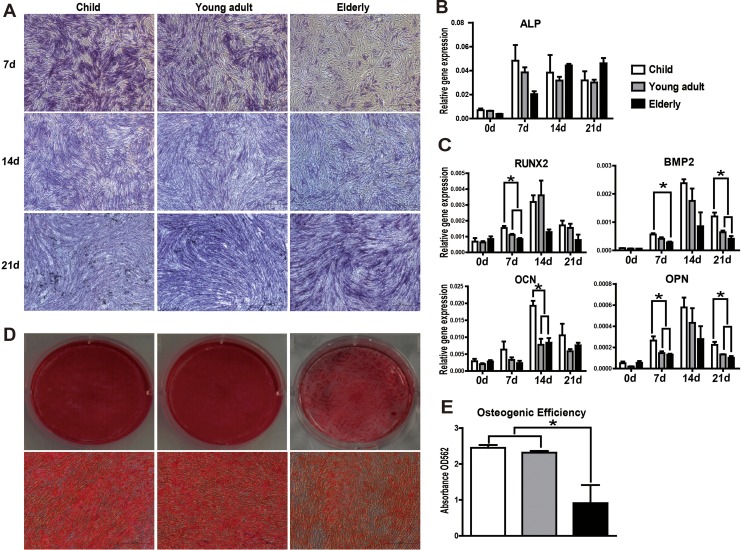
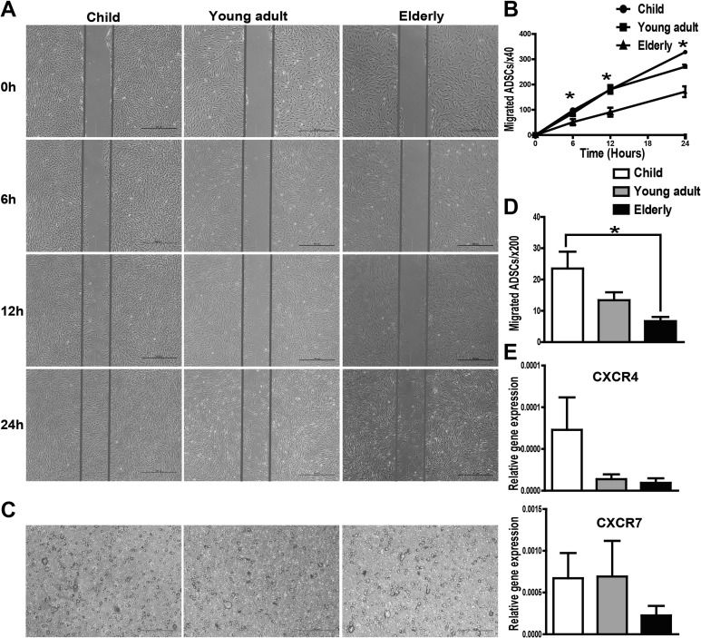
Adipose-derived stem cells (ASCs) can be applied extensively in the clinic because they can be easily isolated and cause less donor-site morbidity; however, their application can be complicated by patient-specific factors, such as age and harvest site. In this study, we systematically evaluated the effects of age on the quantity and quality of human adipose-derived mesenchymal stem cells (hASCs) isolated from excised chest subcutaneous adipose tissue and investigated the underlying molecular mechanism. hASCs were isolated from donors of 3 different age-groups (i.e., child, young adult, and elderly). hASCs are available from individuals across all age-groups and maintain mesenchymal stem cell (MSC) characteristics. However, the increased age of the donors was found to have a significant negative effect on hASCs frequency base on colony-forming unit fibroblasts assay. Moreover, there is a decline in both stromal vascular fraction (SVF) cell yield and the proliferation rate of hASCs with increasing age, although this relationship is not significant. Aging increases cellular senescence, which is manifested as an increase in SA-β-gal-positive cells, increased mitochondrial-specific reactive oxygen species (ROS) production, and the expression of p21 in the elderly. Further, advancing age was found to have a significant negative effect on the adipogenic and osteogenic differentiation potentials of hASCs, particularly at the early and mid-stages of induction, suggesting a slower response to the inducing factors of hASCs from elderly donors. Finally, impaired migration ability was also observed in the elderly group and was determined to be associated with decreased expression of chemokine receptors, such as CXCR4 and CXCR7. Taken together, these results suggest that, while hASCs from different age populations are phenotypically similar, they present major differences at the functional level. When considering potential applications of hASCs in cell-based therapeutic strategies, the negative influence of age on hASC differentiation potential and migration abilities should be taken seriously.

Keywords: aging; cell senescence; differentiation; human adipose-derived mesenchymal stem cells (hASCs); migration.


Conflict of interest statement

Declaration of Conflicting Interests: The author(s) declared no potential conflicts of interest with respect to the research, authorship, and/or publication of this article.

Figures

Fig. 1.
Fig. 1.
Influence of age on stromal vascular fraction (SVF) yield, cell viability, and mesenchymal stem cell (MSC) characteristics. (A) A slight trend indicated that the SVF cell yield decreased with increasing donor age, but no significant difference was found between the different age-groups. (B) Donor age had no effect on human adipose-derived mesenchymal stem cells (hASCs) viability. (C) hASCs from different age-groups showed similar cellular morphologies at passage 0 and 3 (P0 and P3) under phase contrast microscopy (20X). (D) FACS analysis was used to determine the proportion of hASCs expressing MSC surface markers (CD44, CD73, CD90, and CD105). Experiments were carried out in triplicate. Data are expressed as the mean ± standard error of mean (SEM). FACS: fluorescent-activated cell sorting.
Fig. 2.
Fig. 2.
Effect of donor age on human adipose-derived mesenchymal stem cells (hASCs) colony-forming unit fibroblasts (CFU-Fs), proliferation, apoptosis, and cell cycle distribution. (A) CFU-Fs assay showed the number of clone forming cells decreased significantly in elderly group compared with the child group. (B) Donor age had no effect on hASCs proliferation, as evidenced by the MTS assay. (C) qRT-PCR analysis of genes associated with proliferation (c-Jun and c-Fos) showing a decreasing trend with age. (D) Apoptosis was analyzed using a Muse Cell Analyzer, showing no significant difference between the different age-groups. (E) Expression of apoptosis-related genes Bcl-2 and caspase-8 were determined by qRT-PCR and did not appear to be significantly affected by donor age. (F) Cell cycle distribution of hASCs in different age-groups analyzed with a Muse Cell Analyzer. The number of cells in S phase in the elderly group was significantly decreased compared with the other 2 groups. (G) qRT-PCR analysis showed that the expression of CHEK1, a cell cycle-associated gene, was upregulated with increasing age, although the difference was not significant. Experiments were carried out in triplicate. Data are expressed as the mean ± standard error of mean [SEM]. *P < 0.05, **P < 0.01
Fig. 3.
Fig. 3.
Senescence state of human adipose-derived mesenchymal stem cells (hASCs) in different age-groups. (A) Representative pictures of senescence-associated β-galactosidase (SA-β-gal) staining (20× magnification). (B) The number of senescent cells (SA-β-gal-positive) was significantly higher in the elderly group. Positive cells were expressed as a percentage of total hASCs counted. (C) Total intracellular ROS production was measured using the DCFH-DA assay. (D) Quantitative analysis of the DCFH-DA assay revealed a significant increase in ROS production in the elderly group. (E) Representative pictures of MitoSOX Red staining demonstrating that mitochondrial-specific ROS production was increased in the elderly group. (F) Proteasome activity assay showed a decreasing trend with age increasing, although the difference was not significant. (G) Messenger RNA (mRNA) expression levels of SIRT1 and hTERT were determined by qRT-PCR. Although there was no difference in their expression between the different age-groups, both exhibit a decreasing trend with increasing age. (H) qRT-PCR analysis showed that the mRNA expression levels of senescence markers p53, p21, and p16 were increased with increasing donor age. The mRNA levels of RB1 declined in an age-dependent manner, whereas the RB2 expression level in 3 groups was comparable. Only p21 mRNA expression showed significant difference between the child group and the elderly group. The expression of each gene was normalized to the amount of GAPDH RNA to calculate the relative amount of mRNA. All tests were performed in triplicate, and the data are expressed as the mean ± standard error of mean. *P < 0.05.
Fig. 4.
Fig. 4.
Aging influences adipogenic differentiation potential at the early time of induction, while the overall potential to form mature adipocytes is comparable between age-groups. (A) Adipogenesis was confirmed in hASCs by Oil Red O staining. (B) Semi-quantitative detection by colorimetric evaluation of Oil Red O uptake showed no significant differences between the 3 groups. (C, D) Adipogenic differentiation was further confirmed through qRT-PCR analysis of genes related to adipogenic differentiation (PPAR, CEBP-α, FABP4, adiponectin, and LPL). At the early time of induction (day 4), all genes related to adipogenic differentiation were significantly higher in the child group compared to the other 2 groups. (E) The expression of lipid synthesis-related genes (GPDH, FASN, ACC1, and HSL) in hASCs after adipogenic induction for 4, 8, and 12 d. There were no significant differences between the different age-groups. (F) The expression levels of thermogenic markers UCP1 and PGC1α were significantly higher in the child group at the early induction stage. All tests were performed in triplicate, and the data are expressed as the mean ± standard error of mean (SEM). *P < 0.05.
Fig. 5.
Fig. 5.
Osteogenic differentiation diminishes with increasing age. (A) Representative pictures of alkaline phosphatase (ALP) staining (20× magnification). (B, C) Messenger RNA (mRNA) expression levels of osteogenic differentiation-related genes (ALP, RUNX2, BMP2, OCN, and OPN) were determined in human adipose-derived mesenchymal stem cells (hASCs) over the course of osteogenic differentiation by qRT-PCR. hASCs from aged donors had a diminished response to osteogenic inducers relative to that of young donors. (D) Alizarin Red S staining was used to visualize mineralization. Representative images were shown from 3 separate experiments. (E) Quantitative analysis of Alizarin Red S staining in hASCs cultures at day 14 after osteogenic induction revealed a significant decrease in matrix calcification in the elderly group compared to younger donors. All tests were performed in triplicate, and the data are expressed as the mean ± standard error of mean (SEM). *P < 0.05.
Fig. 6.
Fig. 6.
Cell scratch test and transwell assay were used to examine the migration ability of human adipose-derived mesenchymal stem cells (hASCs). (A) Images from the scratch wound migration assay. Healing due to the cellular migration of hASCs was observed over a period of 6 and 24 h following scratch wounding. (B) Quantitative data from the cell scratch healing assay were shown. The migration ability of hASCs in the elderly group was decreased significantly at 6, 12, and 24 h compared to that of the child group. (C) Representative images of cell migration using a transwell assay. (D) Quantitative analysis of cell migration using the transwell assay. The number of migratory cells of the elderly group was reduced compared to that of the child group (within 6 h). (E) The messenger RNA (mRNA) expression levels of chemokine receptors CXCR4 and CXCR7 were decreased in the elderly group. All tests were performed in triplicate, and the data are expressed as the mean ± standard error of mean (SEM). *P < 0.05.

References

    1. Gimble JM, Bunnell BA, Floyd ZE. Prospecting for adipose progenitor cell biomarkers: biopanning for gold with in vivo phage display. Cell Stem Cell. 2011;9(1):1–2. - PubMed
    1. Nicpon J, Marycz K, Grzesiak J. Therapeutic effect of adipose-derived mesenchymal stem cell injection in horses suffering from bone spavin. Pol J Vet Sci. 2013;16(4):753–754. - PubMed
    1. Tzouvelekis A, Paspaliaris V, Koliakos G, Ntolios P, Bouros E, Oikonomou A, Zissimopoulos A, Boussios N, Dardzinski B, Gritzalis D, et al. A prospective, non-randomized, no placebo-controlled, phase Ib clinical trial to study the safety of the adipose derived stromal cells-stromal vascular fraction in idiopathic pulmonary fibrosis. J Transl Med. 2013;11:171. - PMC - PubMed
    1. Murohara T, Shintani S, Kondo K. Autologous adipose-derived regenerative cells for therapeutic angiogenesis. Curr Pharm Des. 2009;15(24):2784–2790. - PubMed
    1. Campisi J. Senescent cells, tumor suppression, and organismal aging: good citizens, bad neighbors. Cell. 2005;120(4):513–522. - PubMed

MeSH terms

LinkOut - more resources