. 2017 Nov 7;8(6):e01263-17.
doi: 10.1128/mBio.01263-17.

Imaging of Cellular Oxidoreductase Activity Suggests Mixotrophic Metabolisms in Thiomargarita spp

Affiliations

Imaging of Cellular Oxidoreductase Activity Suggests Mixotrophic Metabolisms in Thiomargarita spp

Jake V Bailey et al. mBio. .

Abstract

The largest known bacteria, Thiomargarita spp., have yet to be isolated in pure culture, but their large size allows for individual cells to be monitored in time course experiments or to be individually sorted for omics-based investigations. Here we investigated the metabolism of individual cells of Thiomargarita spp. by using a novel application of a tetrazolium-based dye that measures oxidoreductase activity. When coupled with microscopy, staining of the cells with a tetrazolium-formazan dye allows metabolic responses in Thiomargarita spp. to be to be tracked in the absence of observable cell division. Additionally, the metabolic activity of Thiomargarita sp. cells can be differentiated from the metabolism of other microbes in specimens that contain adherent bacteria. The results of our redox dye-based assay suggest that Thiomargarita is the most metabolically versatile under anoxic conditions, where it appears to express cellular oxidoreductase activity in response to the electron donors succinate, acetate, citrate, formate, thiosulfate, H2, and H2S. Under hypoxic conditions, formazan staining results suggest the metabolism of succinate and likely acetate, citrate, and H2S. Cells incubated under oxic conditions showed the weakest formazan staining response, and then only to H2S, citrate, and perhaps succinate. These results provide experimental validation of recent genomic studies of Candidatus Thiomargarita nelsonii that suggest metabolic plasticity and mixotrophic metabolism. The cellular oxidoreductase response of bacteria attached to the exterior of Thiomargarita also supports the possibility of trophic interactions between these largest of known bacteria and attached epibionts.IMPORTANCE The metabolic potential of many microorganisms that cannot be grown in the laboratory is known only from genomic data. Genomes of Thiomargarita spp. suggest that these largest of known bacteria are mixotrophs, combining lithotrophic metabolism with organic carbon degradation. Our use of a redox-sensitive tetrazolium dye to query the metabolism of these bacteria provides an independent line of evidence that corroborates the apparent metabolic plasticity of Thiomargarita observed in recently produced genomes. Finding new cultivation-independent means of testing genomic results is critical to testing genome-derived hypotheses on the metabolic potentials of uncultivated microorganisms.

Keywords: Beggiatoa; Thiomargarita; chemolithotrophy.


Figures

FIG 1
FIG 1
Cellular oxidoreductase activity reduces colorless tetrazolium to a purple formazan product. (A) Initially, Thiomargarita sp. cells incubated in the redox dye are colorless and the medium is light green. (B) Many intact cells under specific treatment conditions, presumably those that are metabolically active, stain a deep purple, and the extracellular medium changes to colorless or light pink, typically within 2 days. Panel B shows cells incubated under anoxic conditions in the presence of succinate. Staining of metabolically active cells was spatially separate and distinct from that of the surrounding sheath material (C). Collapsed or damaged cells at the time of incubation did not exhibit a color change (D), showing exposure to acetate and hydrogen, respectively, under anoxic conditions. Staining of metabolically active cells was distinct in intensity from that of control diatom frustules (E) and control sheath material (F). Upon exposure to H2S, the extracellular medium assumed an orange hue that was readily distinguishable from the purple color change in the Thiomargarita sp. cells (G) and in biofilms of attached epibiont bacteria (arrows). Under anoxic conditions, cells and medium showed the most extensive response, with very little color change observed with no additional electron donor added (H) and a strong staining response with the addition of other substrates such as H2 under anoxic conditions (I and J). In some cases, staining of extracellular bacteria was present as a diffuse stained cloud composed of small cells within the well. A zone characterized by absence of staining and cells in the immediate vicinity of the Thiomargarita cells suggests some sort of inhibition of these small bacteria (arrows in panel K). In other cases, such as exposure to thiosulfate under anoxic conditions, as shown here, stained filamentous epibionts could be observed anchored to the Thiomargarita cell/sheath (L). All images were taken at ×400 magnification. Scale bar in panel A, ~100 μm.
FIG 2
FIG 2
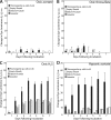
Thiomargarita cells exhibited very little staining response to acetate (A), thiosulfate (B), and other substrates under oxygenated conditions relative to that of controls. However, H2S (C) and citrate (not shown) did induce a statistically significant staining response under oxic conditions. Potentially significant staining was observed under hypoxic conditions in the presence of acetate (D), succinate, citrate, and H2S. Plotted here is the mean change in reciprocal intensity of luminance relative to that at day zero. Error bars indicate the standard error of the mean of the Thiomargarita incubations or controls. a.u., arbitrary units.
FIG 3
FIG 3
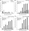
Thiomargarita cells and controls showed a weak response under anoxic conditions with the addition of a substrate that could not be differentiated from controls (A). With the addition of H2 (B), succinate (C), thiosulfate (D), H2S, formate, acetate, citrate, and formate (not shown), a statistically significant staining response was observed under anoxic conditions. Plotted here is the mean change in the reciprocal intensity of luminance relative to that at day zero. Error bars indicate the standard error of the mean of the Thiomargarita incubations or controls. a.u., arbitrary units.

References

    1. Salman V, Bailey JV, Teske A. 2013. Phylogenetic and morphologic complexity of giant sulphur bacteria. Antonie Leeuwenhoek 104:169–186. doi:10.1007/s10482-013-9952-y. - DOI - PubMed
    1. Nelson DC, Wirsen CO, Jannasch HW. 1989. Characterization of large, autotrophic Beggiatoa spp. abundant at hydrothermal vents of the Guaymas Basin. Appl Environ Microbiol 55:2909–2917. - PMC - PubMed
    1. Schulz HN, Brinkhoff T, Ferdelman TG, Mariné MH, Teske A, Jørgensen BB. 1999. Dense populations of a giant sulfur bacterium in Namibian shelf sediments. Science 284:493–495. doi:10.1126/science.284.5413.493. - DOI - PubMed
    1. Gallardo VA. 1977. Large benthic microbial communities in sulphide biota under Peru-Chile subsurface counter current. Nature 286:331–332.
    1. Larkin JM, Strohl WR. 1983. Beggiatoa, Thiothrix and Thioploca. Annu Rev Microbiol 37:341–367. doi:10.1146/annurev.mi.37.100183.002013. - DOI - PubMed

Publication types

LinkOut - more resources