. 2017 Dec 4;149(12):1139-1148.
doi: 10.1085/jgp.201711834. Epub 2017 Nov 7.

Na leak with gating pore properties in hypokalemic periodic paralysis V876E mutant muscle Ca channel

Affiliations

Na leak with gating pore properties in hypokalemic periodic paralysis V876E mutant muscle Ca channel

Clarisse Fuster et al. J Gen Physiol. .

Abstract

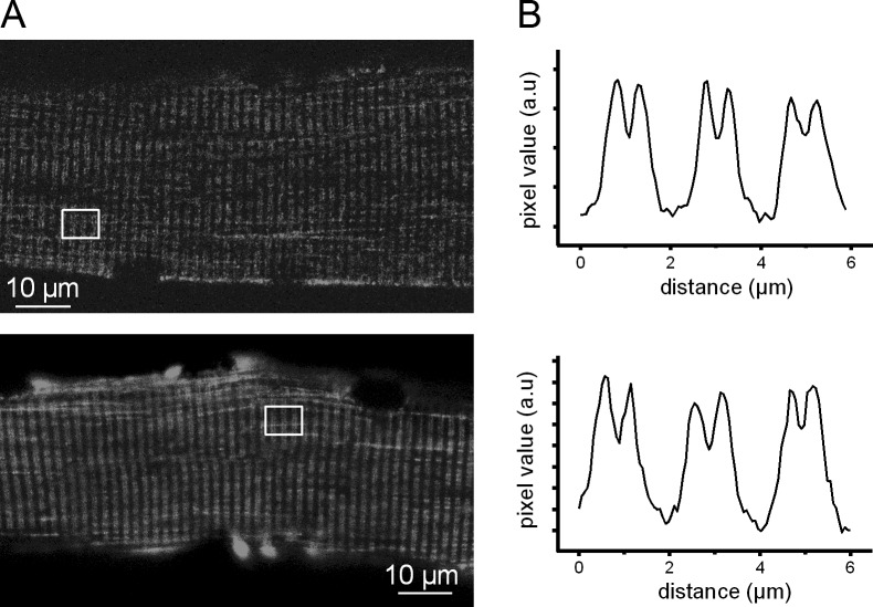
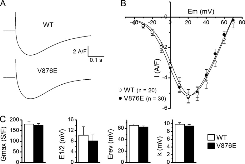
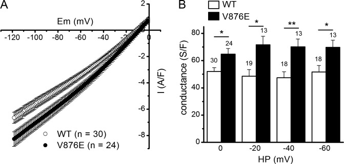
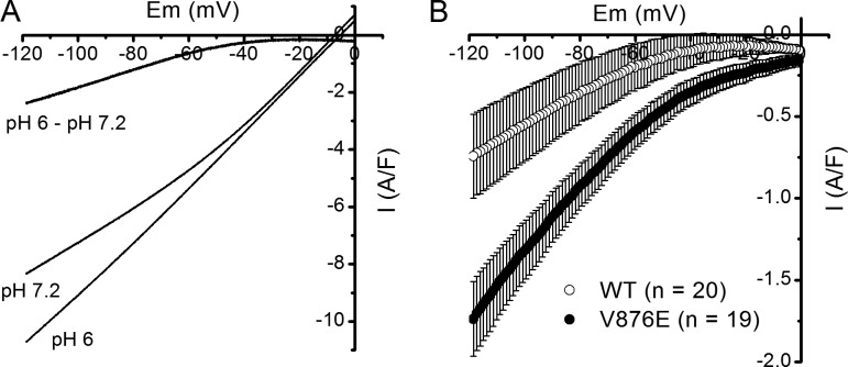
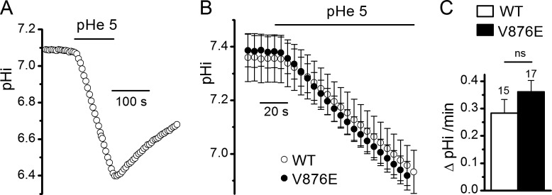
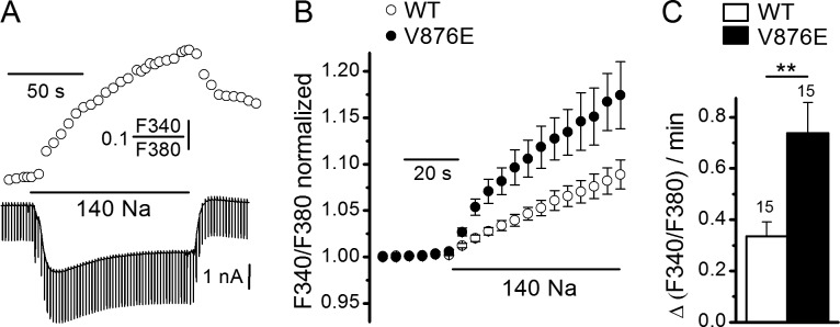
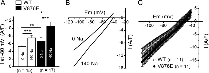
Type 1 hypokalemic periodic paralysis (HypoPP1) is a poorly understood genetic neuromuscular disease characterized by episodic attacks of paralysis associated with low blood K+ The vast majority of HypoPP1 mutations involve the replacement of an arginine by a neutral residue in one of the S4 segments of the α1 subunit of the skeletal muscle voltage-gated Ca2+ channel, which is thought to generate a pathogenic gating pore current. The V876E HypoPP1 mutation has the peculiarity of being located in the S3 segment of domain III, rather than an S4 segment, raising the question of whether such a mutation induces a gating pore current. Here we successfully transfer cDNAs encoding GFP-tagged human wild-type (WT) and V876E HypoPP1 mutant α1 subunits into mouse muscles by electroporation. The expression profile of these WT and V876E channels shows a regular striated pattern, indicative of their localization in the t-tubule membrane. In addition, L-type Ca2+ current properties are the same in V876E and WT fibers. However, in the presence of an external solution containing low-Cl- and lacking Na+ and K+, V876E fibers display an elevated leak current at negative voltages that is increased by external acidification to a higher extent in V876E fibers, suggesting that the leak current is carried by H+ ions. However, in the presence of Tyrode's solution, the rate of change in intracellular pH produced by external acidification was not significantly different in V876E and WT fibers. Simultaneous measurement of intracellular Na+ and current in response to Na+ readmission in the external solution reveals a rate of Na+ influx associated with an inward current, which are both significantly larger in V876E fibers. These data suggest that the V876E mutation generates a gating pore current that carries strong resting Na+ inward currents in physiological conditions that are likely responsible for the severe HypoPP1 symptoms associated with this mutation.


Figures

Figure 1.
Figure 1.
Distribution of GFP-tagged human WT and V876E Cav1.1 in adult mouse skeletal muscle fibers. (A) Confocal images of GFP fluorescence in a fiber expressing WT (top) and V876E (bottom) Cav1.1. (B) Fluorescence intensity profiles from the white box region in the corresponding images in A.
Figure 2.
Figure 2.
L-type voltage-gated Ca2+ currents in WT and V876E fibers. (A) Recordings of L-type currents (top) in a WT and in a V876E fiber in response to depolarizing pulses of 1 s duration to the indicated voltages (bottom). (B) Relationships between the mean peak values of L-type Ca2+ currents and membrane voltage in the two fiber types. (C) Mean of the fitting parameters of current-voltage relationships obtained in each WT and V876E fiber. E1/2, half-activation voltage; Erev, apparent reversal potential; Gmax, maximum conductance; k, steepness factor. Data are given as means ± SEM.
Figure 3.
Figure 3.
Leak currents and leak conductance in WT and V876E fibers. (A) Means and SEM of current densities evoked by voltage ramps applied from a holding potential of 0 mV in WT and R1239H fibers. The number of data points has been reduced for clarity. (B) Mean slope membrane conductance measured between −120 and −80 mV for voltage ramp–evoked membrane currents in the two fiber types from different holding potentials (HP). The number of fibers tested is indicated above each histogram bar. The p-values were 0.009, 0.006, 0.003, and 0.017 at 0, −20, −40, and −60 mV, respectively. Data are given as means ± SEM. *, P < 0.05; **, P < 0.005.
Figure 4.
Figure 4.
Effect of external acidification on leak currents in WT and V876E fibers. (A) Membrane currents evoked by a voltage ramp applied from a holding potential of 0 mV at an external pH of 7.2 and 6, and current difference between pH 6.0 and 7.2 in the same V876E fiber. (B) Means and SEM of current differences between pH 6.0 and pH 7.2 in WT and V876E fibers. The number of data points has been reduced for clarity.
Figure 5.
Figure 5.
Effect of external acidification on pHi in WT and V876E fibers in the presence of an external Tyrode’s solution. (A) Recording of the change in pHi in response to exposition of the cell to an external solution buffered at pH 5.0 in a BCECF-loaded V876E fiber held at −80 mV. (B) Means and SEM of pHi as a function of time in WT and in V876E fibers in response to external acidification (pHe). (C) Mean rate of change in pHi measured during the first minute of exposition of the cell to the external solution buffered at pH 5. The number of fibers tested is indicated above each histogram bar. Data are given as means ± SEM.
Figure 6.
Figure 6.
Effect of external Na+ on SBFI fluorescence ratio and background currents in WT and V876E fibers. (A) Simultaneous recordings of SBFI fluorescence ratio (upper trace) and membrane currents (lower trace) in response to a change of the external solution from a Na+-free NMDG containing solution to a 140 mM Na+–containing solution in a V876E fiber held at −80 mV and stimulated by 50-ms duration voltage pulses given to −90 mV at a frequency of 0.5 Hz. (B) Mean and SEM of SBFI fluorescence ratio as a function of time in response to external Na+ readmission in WT and in V876E fibers. In each cell, values of fluorescence ratio have been normalized to the value of the last data point measured before Na+ readmission. (C) Mean rate of change in SBFI fluorescence ratio measured during the first 30 s of exposure of the cell to external Na+. The number of fibers tested is indicated above each histogram bar. Data are given as means ± SEM. P-value was 0.0043 (**).
Figure 7.
Figure 7.
Effect of external Na+ on leak currents and conductance in WT and V876E fibers. (A) Mean background current intensity measured at −80 mV in the absence and in the presence of external Na+ in WT and V876E fibers. Values were compared with paired tests and unpaired tests for data obtained within the same fiber population and for data obtained between the two fiber populations, respectively. P-values were 0.0001 and 0.0001 for comparison of the means between the Na+-free and the Na+ containing solution in WT and V876E fibers, respectively, and 0.047 for comparison of the means obtained in the presence of the Na+–containing solution in WT and V876E fibers. Data are given as means ± SEM. *, P < 0.05; ***, P < 0.0005. (B) Membrane currents evoked by a voltage ramp applied from a holding potential of −40 mV in the absence and in the presence of external Na+ in the same V876E fiber. (C) Means and SEM of current densities evoked by voltage ramps applied from a holding potential of −40 mV in the presence of external Na+ in WT fibers and V876E fibers. The number of data points has been reduced for clarity.

References

    1. Campos F.V., Chanda B., Roux B., and Bezanilla F.. 2007. Two atomic constraints unambiguously position the S4 segment relative to S1 and S2 segments in the closed state of Shaker K channel. Proc. Natl. Acad. Sci. USA. 104:7904–7909. 10.1073/pnas.0702638104 - DOI - PMC - PubMed
    1. Cannon S.C. 2010. Voltage-sensor mutations in channelopathies of skeletal muscle. J. Physiol. 588:1887–1895. 10.1113/jphysiol.2010.186874 - DOI - PMC - PubMed
    1. Cannon S.C. 2015. Channelopathies of skeletal muscle excitability. Compr. Physiol. 5:761–790. 10.1002/cphy.c140062 - DOI - PMC - PubMed
    1. Catterall W.A. 2011. Voltage-gated calcium channels. Cold Spring Harb. Perspect. Biol. 3:a003947 10.1101/cshperspect.a003947 - DOI - PMC - PubMed
    1. Collet C., Csernoch L., and Jacquemond V.. 2003. Intramembrane charge movement and L-type calcium current in skeletal muscle fibers isolated from control and mdx mice. Biophys. J. 84:251–265. 10.1016/S0006-3495(03)74846-2 - DOI - PMC - PubMed