. 2017 Nov 7;10(504):eaan0852.
doi: 10.1126/scisignal.aan0852.

Aberrant Rac1-cofilin signaling mediates defects in dendritic spines, synaptic function, and sensory perception in fragile X syndrome

Affiliations

Aberrant Rac1-cofilin signaling mediates defects in dendritic spines, synaptic function, and sensory perception in fragile X syndrome

Alexander Pyronneau et al. Sci Signal. .

Abstract

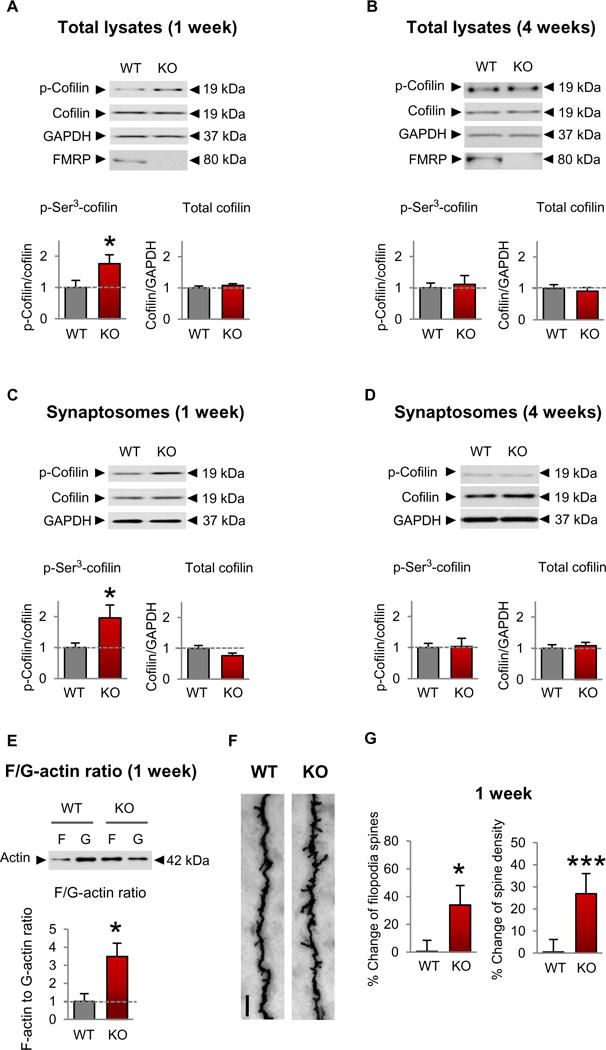
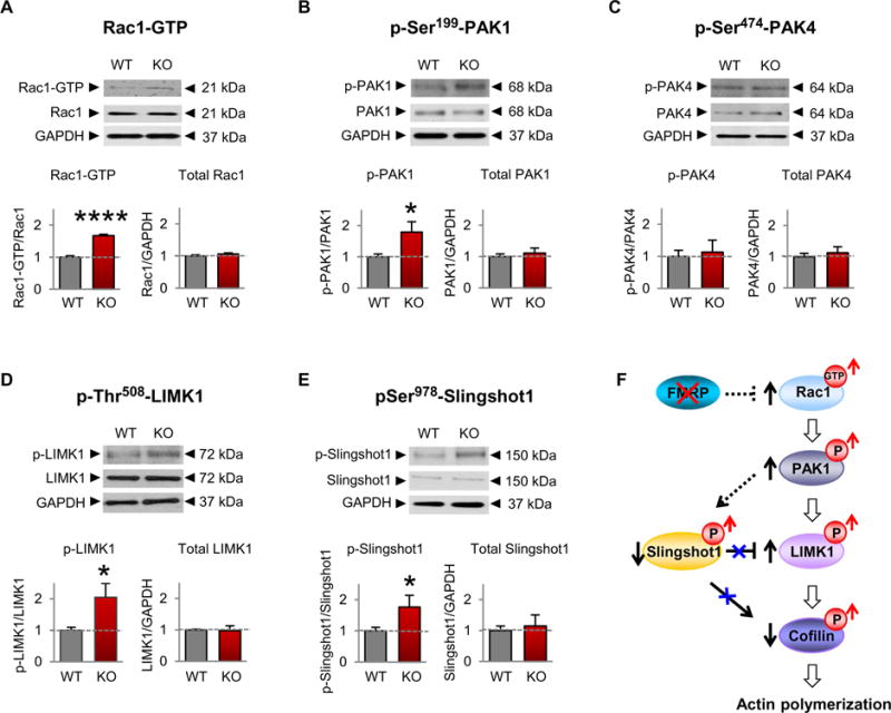
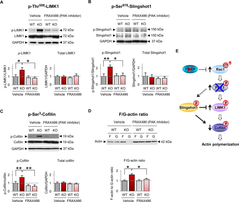
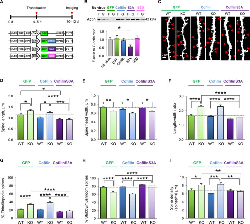
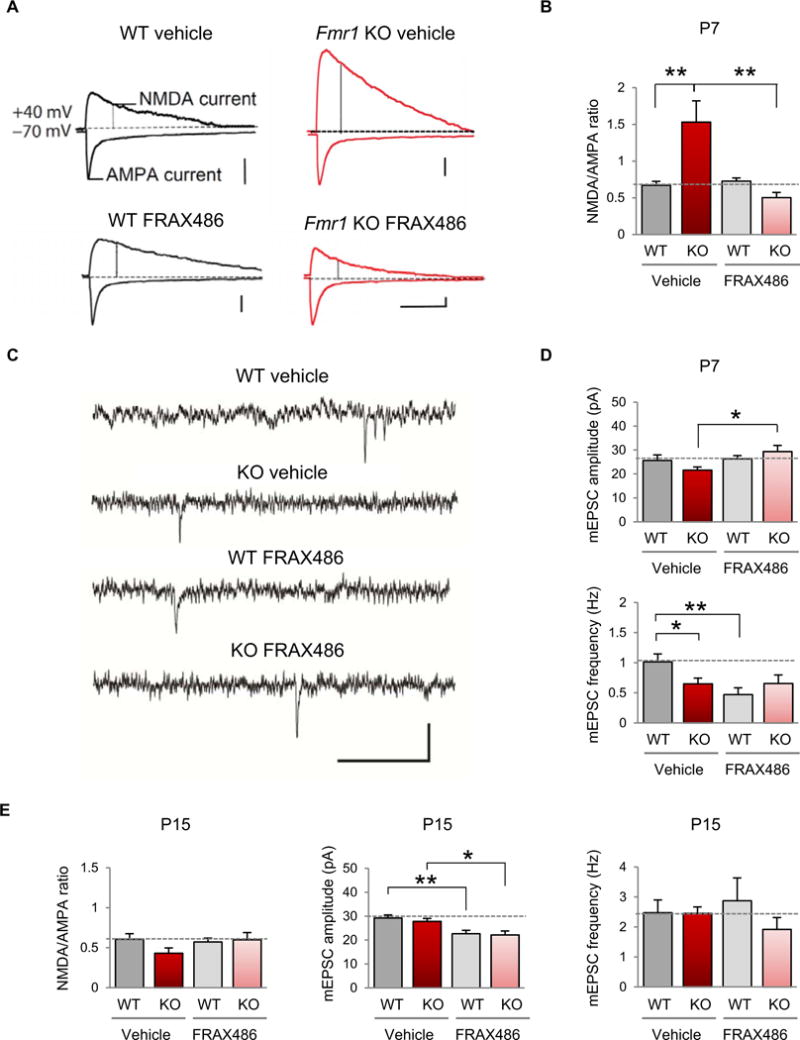
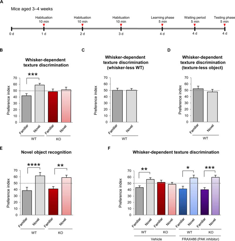
Fragile X syndrome (FXS) is the most common inherited cause of intellectual disabilities and a leading cause of autism. FXS is caused by a trinucleotide expansion in the gene FMR1 on the X chromosome. The neuroanatomical hallmark of FXS is an overabundance of immature dendritic spines, a factor thought to underlie synaptic dysfunction and impaired cognition. We showed that aberrantly increased activity of the Rho GTPase Rac1 inhibited the actin-depolymerizing factor cofilin, a major determinant of dendritic spine structure, and caused disease-associated spine abnormalities in the somatosensory cortex of FXS model mice. Increased cofilin phosphorylation and actin polymerization coincided with abnormal dendritic spines and impaired synaptic maturation. Viral delivery of a constitutively active cofilin mutant (cofilinS3A) into the somatosensory cortex of Fmr1-deficient mice rescued the immature dendritic spine phenotype and increased spine density. Inhibition of the Rac1 effector PAK1 with a small-molecule inhibitor rescued cofilin signaling in FXS mice, indicating a causal relationship between PAK1 and cofilin signaling. PAK1 inhibition rescued synaptic signaling (specifically the synaptic ratio of NMDA/AMPA in layer V pyramidal neurons) and improved sensory processing in FXS mice. These findings suggest a causal relationship between increased Rac1-cofilin signaling, synaptic defects, and impaired sensory processing in FXS and uncover a previously unappreciated role for impaired Rac1-cofilin signaling in the aberrant spine morphology and spine density associated with FXS.


Conflict of interest statement

Competing interests: The authors declare that they have no competing interests.

Figures

Fig. 1
Fig. 1. Increased cofilin phosphorylation and actin polymerization coincide with spine defects in the developing somatosensory cortex of Fmr1 KO mice
(A to D) Representative Western blots (top) and summary data (bottom) assessing the abundance of phosphorylated (p-) and total cofilin in lysates (A and B) and synaptosome fractions (C and D) of the somatosensory cortex of male Fmr1 KO and wild-type (WT) mice at 1 (A and C) and 4 (B and D) weeks of age. WT, n ≥ 12; KO, n ≥ 9 mice. (E) Representative Western blots (top) and summary data (bottom) for F-actin and G-actin in male Fmr1 KO animals and control littermates (WT, n = 5; KO, n = 6). (F) Representative Golgi staining images of the apical dendrites of layer V pyramidal neurons from WT (left) and Fmr1 KO (right) somatosensory cortices isolated from 1-week-old male mice. Scale bar, 5 μm. (G) Summary data showing percent change of immature spines and dendritic spine density (WT, n = 3 animals and 12 dendrites; KO, n = 3 animals and 13 dendrites). Data are means ± SEM. *P < 0.05, ***P < 0.001 (see table S1 for further details of the statistical tests).
Fig. 2
Fig. 2. Rac1 signaling is increased in the somatosensory cortex of FXS mice
(A to E) Representative Western blotting of proteins in the Rac1 signaling pathway in somatosensory lysates (A) or synaptosomes (B to E) from 1-week-old male WT and Fmr1 KO mice (WT, n = 4 to 12;KO, n = 4 to 15). Data are means ± SEM. *P < 0.05, ****P <0.0001. (F) Model depicting a mechanism by which loss of FMRP leads to increased Rac1 signaling, cofilin phosphorylation (a measure of inactivation), and actin polymerization.
Fig. 3
Fig. 3. Inhibition of PAK restores cofilin signaling and actin polymerization in FXS model mice
(A to D) Representative Western blots and summary data for p-Thr508-LIMK (A), p-Ser9780-Slinghot1 (B), p-Ser3-cofilin (C), and the F-actin/G-actin ratio (D) in somatosensory synaptosomes from 1-week-old male WT and Fmr1 KO mice given a single subcutaneous injection of FRAX486 or vehicle [20% (w/v) hydroxypropyl-β-cyclodextrin] 8 hours before sacrifice (WT vehicle, n ≥ 12; KO vehicle, n ≥ 10;WT FRAX486, n ≥ 8;KO FRAX486, n ≥ 9). We note that overall there was no significant difference in total actin abundance in synaptosomes from FRAX486- versus vehicle-treated mice. Data are means ± SEM. *P < 0.05, **P < 0.01. (E) Schematic of Rac-cofilin signaling depicting proposed mechanism by which inhibition of group 1 PAKs (PAK1, PAK2, and PAK3) restores the abundance of p-LIMK1, p-Slingshot1, p-cofilin, and actin polymerization in Fmr1 KO animals.
Fig. 4
Fig. 4. Constitutively active cofilin rescues aberrant spine morphology and density In the somatosensory cortex of young Fmr1 KO mice
(A) Experimental timeline of viral application on cultured neurons or viral delivery and stereotaxic injection into the somatosensory cortex. (B) Representative Western blots and summary data of the F-actin/G-actin ratio in lysates from somatosensory cortical cultures (10 to 12 DIV) either unperturbed or infected with virus expressing GFP, WT cofilin, constitutively active coflinS3A, or phosphomimetic cofilinS3D (n = 13 to 15 culture wells per group from three independent experiments). (C) Representative fluorescent images assessing viral-mediated transduction of apical dendrites of layer V somatosensory cortex pyramidal neurons with GFP (left), GFP-WT cofilin (middle), and GFP-cofilinS3A (right) in male WT and Fmr1 KO mice at P10 to P12. Scale bar, 2 μm. Examples of mature spines (arrowheads) and immature protrusions (arrows) are indicated. (D to I) Summary data of average spine length (D), head width (E), spine length-to-width ratio (LWR) (F), % mature (stubby/mushroom) spines (G), % immature (thin/filopodia) spines (H), and spine density (I) of the samples imaged in (C) (n = 15 to 21 neurons pooled from 6 to 10 animals per group). Data are means ± SEM. *P < 0.05, **P < 0.01, ***P < 0.001, ****P < 0.0001.
Fig. 5
Fig. 5. Inhibition of PAK corrects functional synaptic deficits in layer V of the somatosensory cortex
(A) Representative evoked EPSC recordings from male WT (left) and Fmr1 KO (right) mice, either vehicle-treated (top) or FRAX486-treated (bottom) at P7. AMPAR-mediated EPSCs were measured as the peak current at −70 mV, and the NMDA component was measured by depolarizing the cell to +40 mV and measuring the current 60 ms after the onset of the outward current in the presence of 50 μM picrotoxin. Calibration: 100 ms, 50 pA. (B) Summary data of the NMDA/AMPA ratio in all recordings (WT vehicle, n = 13; Fmr1 KO vehicle, n = 11; WT FRAX486, n = 11; Fmr1 KO FRAX486, n = 6). (C) Representative traces of mEPSC recordings from male WT and Fmr1 KO animals, vehicle- or FRAX486-treated at P7. Calibration: 500 ms, 50 pA. (D) Summary data of mEPSC amplitude and frequency in all recordings (WT vehicle, n = 10; Fmr1 KO vehicle, n = 10; WT FRAX486, n = 11; Fmr1 KO FRAX486, n = 9). (E) Summary data for all recordings at P15 (NMDA/AMPA ratio: WT vehicle, n = 15; Fmr1 KO vehicle, n = 7; WT FRAX486, n =11; Fmr1 KO FRAX486, n = 10; mEPSC amplitude and frequency: WT vehicle, n = 18; Fmr1 KO, n = 7; WT FRAX486, n = 16; Fmr1 KO FRAX486, n = 12). Data are means ± SEM. *P < 0.05, **P < 0.01.
Fig. 6
Fig. 6. Inhibition of PAK corrects impaired sensory processing in FXS model mice
(A) Timeline of the whisker-dependent texture discrimination task. (B) Discrimination of a novel versus familiar texture in WT and Fmr1 KO mice, as assessed by a preference index at chance level indicating impaired sensory processing (WT, n = 13; KO, n = 7). (C and D) Discrimination of a novel texture versus a familiar texture WT mice subjected to (C) whisker trimming (n = 10 animals) or (D) a texture-less object (n = 7 animals), indicating the necessity for an intact mystacial vibrissae to discriminate textures. (E) Novel object recognition task in WT and Fmr1 KO mice (WT, n = 7; KO, n = 8 animals). (F) Sensory processing, as assessed by the ability to discriminate a novel texture versus a familiar texture, in Fmr1 KO animals subjected to chronic administration of FRAX486 (once at P7, once at P14, and once at 3 to 4 weeks of age 24 hours before testing) (WT vehicle, n = 15; KO vehicle, n = 9; WT FRAX486, n = 10;KO FRAX486, n =10). Data are means ± SEM. *P < 0.05, **P < 0.01, ***P < 0.001.

References

    1. Bagni C, Tassone F, Neri G, Hagerman R. Fragile X syndrome: Causes, diagnosis, mechanisms, and therapeutics. J Clin Invest. 2012;122:4314–4322. - PMC - PubMed
    1. Bhakar AL, Dölen G, Bear MF. The pathophysiology of fragile X (and what it teaches us about synapses) Annu Rev Neurosci. 2012;35:417–443. - PMC - PubMed
    1. Huber KM, Klann E, Costa-Mattioli EM, Zukin RS. Dysregulation of mammalian target of rapamycin signaling in mouse models of autism. J Neurosci. 2015;41:13836–13842. - PMC - PubMed
    1. Gross C, Hoffmann A, Bassell GJ, Berry-Kravis EM. Therapeutic strategies in fragile X syndrome: From bench to bedside and back. Neurotherapeutics. 2015;12:584–608. - PMC - PubMed
    1. Lüscher C, Huber KM. Group 1 mGluR-dependent synaptic long-term depression: Mechanisms and implications for circuitry and disease. Neuron. 2010;65:445–459. - PMC - PubMed

MeSH terms