Review
. 2017 Dec 4;216(12):3903-3916.
doi: 10.1083/jcb.201702150. Epub 2017 Nov 7.

Regulation and dysregulation of axon infrastructure by myelinating glia

Affiliations
Review

Regulation and dysregulation of axon infrastructure by myelinating glia

Simon Pan et al. J Cell Biol. .

Abstract

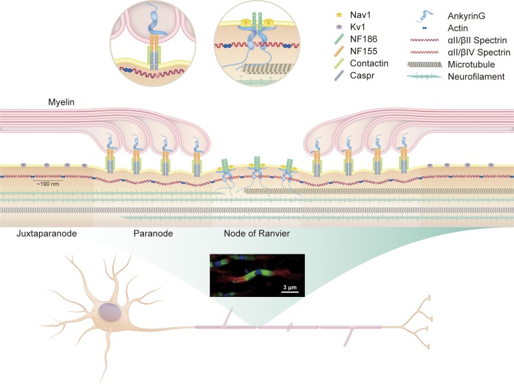
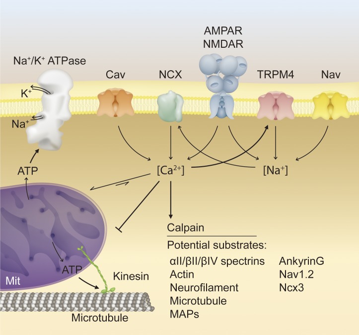
Axon loss and neurodegeneration constitute clinically debilitating sequelae in demyelinating diseases such as multiple sclerosis, but the underlying mechanisms of secondary degeneration are not well understood. Myelinating glia play a fundamental role in promoting the maturation of the axon cytoskeleton, regulating axon trafficking parameters, and imposing architectural rearrangements such as the nodes of Ranvier and their associated molecular domains. In the setting of demyelination, these changes may be reversed or persist as maladaptive features, leading to axon degeneration. In this review, we consider recent insights into axon-glial interactions during development and disease to propose that disruption of the cytoskeleton, nodal architecture, and other components of axon infrastructure is a potential mediator of pathophysiological damage after demyelination.


Figures

Figure 1.
Figure 1.
Architecture of axonal cytoskeleton and polarized molecular domains. Myelination induces architectural rearrangement of the axon into polarized molecular domains. The nodes of Ranvier are short, unmyelinated segments that contain clusters of voltage-gated sodium channels. Flanking the nodes are the paranodes, junctions between noncompacted paranodal myelin loops and the underlying axolemma. Distal to the paranodes are the juxtaparanodes, which contain voltage-gated potassium channels. The fluorescent micrograph of optic nerve axons illustrates the distinct localization of potassium channels (red, juxtaparanode), Caspr (green, paranode), and βIV spectrin (blue, node). The axon cytoskeleton facilitates structural integrity and molecular organization and acts as a conduit for axon transport. It consists primarily of neurofilaments, microtubules, actins, and spectrins. Actins and spectrins assemble into a repeating lattice with structural periodicity of 180 to 190 nm. At nodes, ankyrin adaptor proteins anchor nodal constituents such as voltage-gated ion channels to the underlying actin–spectrin cytoskeleton. At paranodes, protein 4.1B (not depicted), anchors the NF155–Caspr–Contactin complex to the underlying actin–spectrin cytoskeleton.
Figure 2.
Figure 2.
Proposed molecular mechanisms of secondary axon degeneration. Multiple pathological mechanisms converge synergistically on axon degeneration. In a demyelinated axon, disassembly of nodal architecture and its underlying cytoskeleton may lead to voltage-gated sodium channel declustering, increasing sodium ion influx and the metabolic demand of action potential conduction. Impaired axon transport, increased metabolic load, and excitotoxic stress may lead to failure of the Na+/K+ ATPase, reversal of the NCX, and entry of calcium into the axon. Increased axoplasmic calcium leads to activation of the calcium-dependent protease calpain, which cleaves many putative substrates crucial to axon function, including those associated with the axon cytoskeleton or transport machinery. Overloading of mitochondrial calcium buffering capacity and impairment of mitochondrial transport via disruption of motor proteins and microtubules inhibits ATP production and exacerbates metabolic impairment. Excitotoxic stress further contributes to increased sodium and calcium influx, transmitted through various ion channels. NMDAR, N-Methyl-d-aspartate receptor. AMPAR, α-amino-3-hydroxy-5-methyl-4-isoxazolepropionic acid receptor; Cav, voltage-gated calcium channel; MAP, microtubule-associated protein; Mit, mitochondria; Nav, voltage-gated sodium channel.

References

    1. Amor V., Zhang C., Vainshtein A., Zhang A., Zollinger D.R., Eshed-Eisenbach Y., Brophy P.J., Rasband M.N., and Peles E.. 2017. The paranodal cytoskeleton clusters Na(+) channels at nodes of Ranvier. eLife. 6:e21392 10.7554/eLife.21392 - DOI - PMC - PubMed
    1. Armstrong R., Toews A.D., and Morell P.. 1987. Axonal transport through nodes of Ranvier. Brain Res. 412:196–199. 10.1016/0006-8993(87)91461-2 - DOI - PubMed
    1. Arroyo E.J., Sirkowski E.E., Chitale R., and Scherer S.S.. 2004. Acute demyelination disrupts the molecular organization of peripheral nervous system nodes. J. Comp. Neurol. 479:424–434. 10.1002/cne.20321 - DOI - PubMed
    1. Bano D., Young K.W., Guerin C.J., Lefeuvre R., Rothwell N.J., Naldini L., Rizzuto R., Carafoli E., and Nicotera P.. 2005. Cleavage of the plasma membrane Na+/Ca2+ exchanger in excitotoxicity. Cell. 120:275–285. 10.1016/j.cell.2004.11.049 - DOI - PubMed
    1. Bechtold D.A., Miller S.J., Dawson A.C., Sun Y., Kapoor R., Berry D., and Smith K.J.. 2006. Axonal protection achieved in a model of multiple sclerosis using lamotrigine. J. Neurol. 253:1542–1551. 10.1007/s00415-006-0204-1 - DOI - PubMed

Publication types

MeSH terms