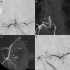
Angiographic and Clinical Characteristics of Thoracolumbar Spinal Epidural and Dural Arteriovenous Fistulas
- PMID: 29114089
- PMCID: PMC5704665
- DOI: 10.1161/STROKEAHA.117.019131
Angiographic and Clinical Characteristics of Thoracolumbar Spinal Epidural and Dural Arteriovenous Fistulas
Abstract
Background and purpose: The purpose of this study is to compare the angiographic and clinical characteristics of spinal epidural arteriovenous fistulas (SEAVFs) and spinal dural arteriovenous fistulas (SDAVFs) of the thoracolumbar spine.
Methods: A total of 168 cases diagnosed as spinal dural or extradural arteriovenous fistulas of the thoracolumbar spine were collected from 31 centers. Angiography and clinical findings, including symptoms, sex, and history of spinal surgery/trauma, were retrospectively reviewed. Angiographic images were evaluated, with a special interest in spinal levels, feeders, shunt points, a shunted epidural pouch and its location, and drainage pattern, by 6 readers to reach a consensus.
Results: The consensus diagnoses by the 6 readers were SDAVFs in 108 cases, SEAVFs in 59 cases, and paravertebral arteriovenous fistulas in 1 case. Twenty-nine of 59 cases (49%) of SEAVFs were incorrectly diagnosed as SDAVFs at the individual centers. The thoracic spine was involved in SDAVFs (87%) more often than SEAVFs (17%). Both types of arteriovenous fistulas were predominant in men (82% and 73%) and frequently showed progressive myelopathy (97% and 92%). A history of spinal injury/surgery was more frequently found in SEAVFs (36%) than in SDAVFs (12%; P=0.001). The shunt points of SDAVFs were medial to the medial interpedicle line in 77%, suggesting that SDAVFs commonly shunt to the bridging vein. All SEAVFs formed an epidural shunted pouch, which was frequently located in the ventral epidural space (88%) and drained into the perimedullary vein (75%), the paravertebral veins (10%), or both (15%).
Conclusions: SDAVFs and SEAVFs showed similar symptoms and male predominance. SDAVFs frequently involve the thoracic spine and shunt into the bridging vein. SEAVFs frequently involve the lumbar spine and form a shunted pouch in the ventral epidural space draining into the perimedullary vein.
Keywords: angiography; arteriovenous malformations; drainage; male; spinal injuries.
© 2017 The Authors.
Figures


Similar articles
-
Clinical and Imaging Characteristics of Spinal Dural Arteriovenous Fistulas and Spinal Epidural Arteriovenous Fistulas.Neurosurgery. 2021 Feb 16;88(3):666-673. doi: 10.1093/neuros/nyaa492. Neurosurgery. 2021. PMID: 33428765
-
Intramedullary Hemorrhage Caused by Lumbosacral Epidural Arteriovenous Fistula with Dual Retrograde Perimedullary Venous Draining Routes: A Case Report and Review of the Literature.World Neurosurg. 2020 Nov;143:295-307. doi: 10.1016/j.wneu.2020.08.025. Epub 2020 Aug 10. World Neurosurg. 2020. PMID: 32791223
-
Accuracy and pitfalls of multidetector-row computed tomography in detecting spinal dural arteriovenous fistulas.J Neurosurg Spine. 2010 Mar;12(3):243-8. doi: 10.3171/2009.9.SPINE0971. J Neurosurg Spine. 2010. PMID: 20192621
-
Presentation and outcomes of patients with thoracic and lumbosacral spinal epidural arteriovenous fistulas: a systematic review and meta-analysis.J Neurointerv Surg. 2019 Jan;11(1):95-98. doi: 10.1136/neurintsurg-2018-014203. Epub 2018 Aug 30. J Neurointerv Surg. 2019. PMID: 30166334
-
Spinal ventral epidural arteriovenous fistulas of the lumbar spine: angioarchitecture and endovascular treatment.Neuroradiology. 2013 Feb;55(3):327-36. doi: 10.1007/s00234-012-1130-9. Epub 2013 Jan 11. Neuroradiology. 2013. PMID: 23306215 Free PMC article. Review.
Cited by
-
Spinal dural and epidural arteriovenous fistula: Recurrence rate after surgical and endovascular treatment.Front Surg. 2023 Apr 4;10:1148968. doi: 10.3389/fsurg.2023.1148968. eCollection 2023. Front Surg. 2023. PMID: 37082364 Free PMC article.
-
Spinal Cord Infarction after Transcatheter Embolization of Pelvic Arteriovenous Malformation.Ann Vasc Dis. 2020 Jun 25;13(2):176-179. doi: 10.3400/avd.cr.19-00111. Ann Vasc Dis. 2020. PMID: 32595795 Free PMC article.
-
Rupture of a Spinal Dural Arteriovenous Fistula as a Differential Diagnosis of a Coronary Syndrome: Case Report.Neurosurg Pract. 2023 Aug 11;4(3):e00050. doi: 10.1227/neuprac.0000000000000050. eCollection 2023 Sep. Neurosurg Pract. 2023. PMID: 39958797 Free PMC article.
-
Freiburg Neuropathology Case Conference: Gait Ataxia, Segmental Hypoesthesia, and Combined Incontinence in a 57-Years-Old Patient.Clin Neuroradiol. 2025 Jun;35(2):417-425. doi: 10.1007/s00062-025-01528-1. Epub 2025 May 13. Clin Neuroradiol. 2025. PMID: 40358711 Free PMC article. No abstract available.
-
Spinal Dural Arteriovenous Fistula: The Missing-Piece Sign.Ochsner J. 2022 Spring;22(1):10-14. doi: 10.31486/toj.21.0110. Ochsner J. 2022. PMID: 35355649 Free PMC article. No abstract available.
References
-
- Fugate JE, Lanzino G, Rabinstein AA. Clinical presentation and prognostic factors of spinal dural arteriovenous fistulas: an overview. Neurosurg Focus. 2012;32:E17. doi: 10.3171/2012.1.FOCUS11376. - PubMed
-
- Marcus J, Schwarz J, Singh IP, Sigounas D, Knopman J, Gobin YP, et al. Spinal dural arteriovenous fistulas: a review. Curr Atheroscler Rep. 2013;15:335. doi: 10.1007/s11883-013-0335-7. - PubMed
-
- Geibprasert S, Pereira V, Krings T, Jiarakongmun P, Toulgoat F, Pongpech S, et al. Dural arteriovenous shunts: a new classification of craniospinal epidural venous anatomical bases and clinical correlations. Stroke. 2008;39:2783–2794. doi: 10.1161/STROKEAHA.108.516757. - PubMed
Publication types
MeSH terms
LinkOut - more resources
Full Text Sources
Other Literature Sources

