Concurrent Multilevel Spinal Intra-medullary with Extensive Intracranial Tuberculomas: A Rare Case Report
- PMID: 29114310
- PMCID: PMC5652122
- DOI: 10.4103/1793-5482.185073
Concurrent Multilevel Spinal Intra-medullary with Extensive Intracranial Tuberculomas: A Rare Case Report
Abstract
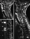
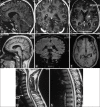
Disseminated tuberculomas in the brain and spinal cord are rare. To the best of our knowledge, only nine cases of spinal intra-medullary tuberculomas with cranial involvement have been reported till date. However, involvement of all levels in the spinal cord, brain stem with pan lobar involvement of the cerebrum and cerebellum has not been reported so far. We present such a case of a 12-year-old boy with history of pulmonary tuberculosis, who presented with gradual onset of quadriparesis and generalized seizures. We have discussed the unusual clinical presentation and the temporal changes in magnetic resonance imaging features along with clinical response to treatment. In cases reported so far, the plan of surgical versus medical management has been opted for variably, in cases of spinal intra-medullary involvement with acute neurological deficit. The decision is even more difficult in multilevel spinal intra-medullary tuberculomas. Our patient showed good clinico-radiological improvement with medical management.
Keywords: Central nervous system infection; concomitant cranio-spinal tuberculoma; intracranial tuberculoma; spinal tuberculoma; tuberculoma.
Conflict of interest statement
There are no conflicts of interest.
Figures


Similar articles
-
Multiple tuberculomas in the brain and spinal cord: a case report.Spine (Phila Pa 1976). 2003 Dec 1;28(23):E499-502. doi: 10.1097/01.BRS.0000099114.40764.46. Spine (Phila Pa 1976). 2003. PMID: 14652486
-
Concurrent multiple intracranial and intramedullary conus tuberculoma: A rare case report.Asian J Neurosurg. 2017 Apr-Jun;12(2):331-333. doi: 10.4103/1793-5482.143461. Asian J Neurosurg. 2017. PMID: 28484568 Free PMC article.
-
Multiple Intracranial Tuberculomas with an Intra-medullary Spinal Cord Tuberculoma in a Pediatric Patient.Cureus. 2020 Mar 12;12(3):e7248. doi: 10.7759/cureus.7248. Cureus. 2020. PMID: 32292663 Free PMC article.
-
Spinal Intramedullary Tuberculosis with Concurrent Supra- and Infratentorial Intracranial Disease in a 9-Month-Old Boy: Case Report and Comprehensive Review of the Literature.World Neurosurg. 2017 Oct;106:37-45. doi: 10.1016/j.wneu.2017.05.069. Epub 2017 May 19. World Neurosurg. 2017. PMID: 28532916 Review.
-
[Extramedullary intradural tuberculosis: a case report and review of the literature].Rev Neurol. 2018 Jan 1;66(1):21-24. Rev Neurol. 2018. PMID: 29251339 Review. Spanish.
Cited by
-
Intracranial and Spinal Tuberculosis: A Rare Entity.Cureus. 2021 Dec 28;13(12):e20787. doi: 10.7759/cureus.20787. eCollection 2021 Dec. Cureus. 2021. PMID: 35111471 Free PMC article.
-
Extensive perivascular dissemination of cerebral miliary tuberculomas: a case report and review of the literature.Acta Radiol Open. 2018 Dec 11;7(12):2058460118817918. doi: 10.1177/2058460118817918. eCollection 2018 Dec. Acta Radiol Open. 2018. PMID: 30559977 Free PMC article.
-
Pott's Disease with Incidentally Discovered Multiple Brain Tuberculomas in a Previously Healthy 10-Year-Old Girl.Case Rep Infect Dis. 2021 Apr 20;2021:5552351. doi: 10.1155/2021/5552351. eCollection 2021. Case Rep Infect Dis. 2021. PMID: 33996161 Free PMC article.
-
Coexisting Spinal Intramedullary and Intracranial Tuberculomas in an Immunocompetent Child.J Pediatr Neurosci. 2019 Jul-Sep;14(3):143-147. doi: 10.4103/jpn.JPN_12_19. Epub 2019 Sep 27. J Pediatr Neurosci. 2019. PMID: 31649775 Free PMC article.
-
Concurrent intracranial tuberculomas and spinal tuberculosis with multifocal abscesses: a rare case report.Ann Med Surg (Lond). 2025 Feb 7;87(3):1724-1728. doi: 10.1097/MS9.0000000000003024. eCollection 2025 Mar. Ann Med Surg (Lond). 2025. PMID: 40213229 Free PMC article.
References
-
- MacDonnell AH, Baird RW, Bronze MS. Intramedullary tuberculomas of the spinal cord: Case report and review. Rev Infect Dis. 1990;12:432–9. - PubMed
-
- Shen WC, Cheng TY, Lee SK, Ho YJ, Lee KR. Disseminated tuberculomas in spinal cord and brain demonstrated by MRI with gadolinium-DTPA. Neuroradiology. 1993;35:213–5. - PubMed
-
- Huang CR, Lui CC, Chang WN, Wu HS, Chen HJ. Neuroimages of disseminated neurotuberculosis: Report of one case. Clin Imaging. 1999;23:218–22. - PubMed
-
- Yen HL, Lee RJ, Lin JW, Chen HJ. Multiple tuberculomas in the brain and spinal cord: A case report. Spine (Phila Pa 1976) 2003;28:E499–502. - PubMed
Publication types
LinkOut - more resources
Full Text Sources
Other Literature Sources

