Review
. 2017 Dec;18(12):728-742.
doi: 10.1038/nrm.2017.108. Epub 2017 Nov 8.

Mechanical forces direct stem cell behaviour in development and regeneration

Affiliations
Review

Mechanical forces direct stem cell behaviour in development and regeneration

Kyle H Vining et al. Nat Rev Mol Cell Biol. 2017 Dec.

Abstract

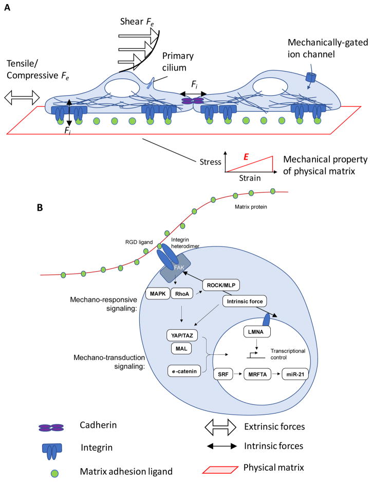
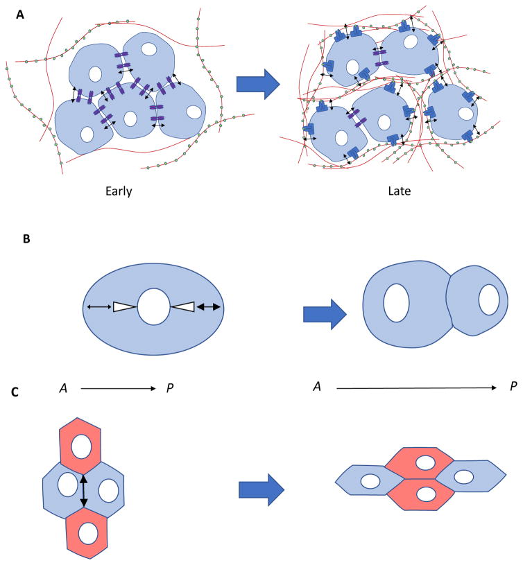
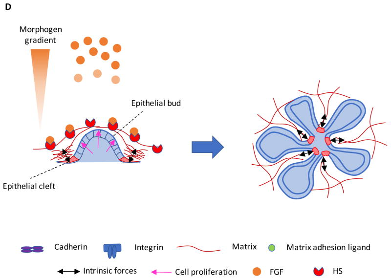
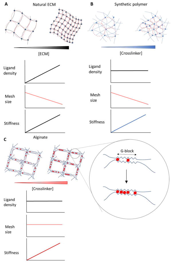
Stem cells and their local microenvironment, or niche, communicate through mechanical cues to regulate cell fate and cell behaviour and to guide developmental processes. During embryonic development, mechanical forces are involved in patterning and organogenesis. The physical environment of pluripotent stem cells regulates their self-renewal and differentiation. Mechanical and physical cues are also important in adult tissues, where adult stem cells require physical interactions with the extracellular matrix to maintain their potency. In vitro, synthetic models of the stem cell niche can be used to precisely control and manipulate the biophysical and biochemical properties of the stem cell microenvironment and to examine how the mode and magnitude of mechanical cues, such as matrix stiffness or applied forces, direct stem cell differentiation and function. Fundamental insights into the mechanobiology of stem cells also inform the design of artificial niches to support stem cells for regenerative therapies.


Conflict of interest statement

COMPETING INTERESTS STATEMENT

The authors declare no competing interests.

Figures

Figure 1
Figure 1
Stem cells exert forces and are subject to external forces, which regulate their intracellular signaling pathways. A) Intrinsic, or cell-generated forces, (Fi) are generated intracellularly and transferred to other cells through cell-cell junctions, like cadherin receptors, or via traction on extracellular matrix (ECM) adhesion ligands that are bound to integrin receptors. Cells are directly coupled by cell-cell junctions, which link the intrinsic forces of one cell to the cytoskeleton of another. Indirect mechanical coupling between cells occurs by intrinsic forces exerted on an ECM, to which two or more cells are adhered. Physical properties, e.g., elastic modulus, E, of the ECM, govern how mechanical cues are transduced. Extrinsic forces (Fe) are externally applied by shear or tension/compression on cells, and can be sensed by mechanically-gated ion channels, changes in receptor-ligand binding, deformation of the cytoskeleton, and the primary cilium. The cytoskeleton generates and transfers forces from membrane proteins to intracellular structures, like the nucleus. B) ECM and intracellular pathways are biochemically coupled by mechanotransduction pathways. Matrix mechanical resistance to intrinsic forces regulates the stability of focal adhesion complexes that contain focal adhesion kinase (FAK), which phosphorylates and activates mechano-responsive signaling elements, such as mitogen-associated protein kinase (MAPK) and Rho kinase (RhoA). RhoA regulates mechanical feedback by activating ROCK, which phosphorylates myosin light chain (MLP) to generate actomyosin forces. For example, RhoA phosphorylates ROCK, which activates non-muscle myosin II contractility by phosphorylation of myosin light chain (MLP), and upregulates mechano-transduction pathways, such as MAL, a G-actin-binding coactivator of serum response factor (SRF),. Yes-associated protein (YAP), and transcriptional coactivator with PDZ-binding motif (TAZ), which induce nuclear transcription via activation and nuclear translocation. Nuclear signaling pathways, such as serum response factor (SRF) and myocardin-related transcription factor-A (MRFTA), regulate transcription of mechanoresponsive genes. For example, mechanics-dependent expression of miR-21 regulates fibrogenic behavior. Wnt activation also promotes activation and nuclear translocation of YAP/TAZ and beta-catenin. Mechanical forces are also physically directly coupled to the nucleus via lamina proteins, such as lamin A (LMNA), which can affect chromatin structure and epigenetic regulation of transcription.
Figure 2
Figure 2
Mechanobiology in the developmental niche. A) While development progresses, intrinsic forces exerted by cells transition from largely cell-cell to more cell-matrix transmission as matrix content in tissues increases. B) Higher astral tension (white triangles) on the posterior axis (right side, bold arrow) of dividing cells is generated by cortical tension and microtubule polymerization, which results in asymmetry in cell size after division. C) Cell-cell intrinsic forces in early development modify the pattern of embryonic epithelial adhesion and intercalation, which results in elongation of the anterior- (A) posterior (P) axis. D) During epithelial branching morphogenesis of the fetal submandibular salivary gland, cells exert intrinsic actomyosin contractility and traction forces on the extracellular matrix (ECM) (red), which assembles at a clefting region and promotes cell proliferation (pink arrow) in the budding region. ECM contains domains of heparan sulfate (HS) that bind FGF growth factors to promote epithelial bud elongation by differentially increasing their local concentration. Thus, concerted biochemical and mechanical cues work together to generate proper organ form.
Figure 2
Figure 2
Mechanobiology in the developmental niche. A) While development progresses, intrinsic forces exerted by cells transition from largely cell-cell to more cell-matrix transmission as matrix content in tissues increases. B) Higher astral tension (white triangles) on the posterior axis (right side, bold arrow) of dividing cells is generated by cortical tension and microtubule polymerization, which results in asymmetry in cell size after division. C) Cell-cell intrinsic forces in early development modify the pattern of embryonic epithelial adhesion and intercalation, which results in elongation of the anterior- (A) posterior (P) axis. D) During epithelial branching morphogenesis of the fetal submandibular salivary gland, cells exert intrinsic actomyosin contractility and traction forces on the extracellular matrix (ECM) (red), which assembles at a clefting region and promotes cell proliferation (pink arrow) in the budding region. ECM contains domains of heparan sulfate (HS) that bind FGF growth factors to promote epithelial bud elongation by differentially increasing their local concentration. Thus, concerted biochemical and mechanical cues work together to generate proper organ form.
Figure 3
Figure 3
Material systems to study stem cell mechanobiology. When engineering a synthetic niche, alterations in the overall polymer concentration may change the density of adhesion ligands, while changing crosslinking without altering the polymer content may vary the network mesh size (spacing between crosslinks), which can affect how molecules diffuse through the network. A) Artificial niches fabricated from naturally-derived ECM typically manipulate stiffness by altering the concentration of the matrix proteins, which increases ligand density and decreases mesh size in parallel. B) Synthetic polymer systems can offer independent control of stiffness and ligand density, by maintaining a constant polymer concentration while altering the crosslink density. However, the mesh size is altered in parallel. C) Matrices formed from alginate polymers can be crosslinked to various extents while maintaining constant ligand density and mesh size, and thus enable one to independently examine how matrix stiffness affects stem cells. (Inset) Crosslinking in this system occurs via cooperative sharing of divalent cations (red) in blocks of one type of sugar residue (G-block) on the chains, and increases in the number of crosslink sites occupied in the aligned blocks do not alter the architecture of the chains. D) Alginate polymer molecular weight (MW) can be used to control the viscoelasticity of an ionically-crosslinked alginate network. Low MW alginate (red arrow and box) forms into a network with less physical entanglement and overlap of the alginate chains. High MW alginate (purple arrow and box) has higher chain entanglement and overlap (shaded blue region), which decreases the ability of the polymer network dissipate stress. E) The low MW network (red line) is more viscous, shown by its rapid relaxation of stress while a constant strain is applied. The high MW network (purple line) dissipates stress more slowly due to more physical entanglement and overlap. The covalently-crosslinked network (black line) is more elastic than the viscoelastic reversibly-crosslinked alginate, and does not significantly dissipate stress over time.
Figure 3
Figure 3
Material systems to study stem cell mechanobiology. When engineering a synthetic niche, alterations in the overall polymer concentration may change the density of adhesion ligands, while changing crosslinking without altering the polymer content may vary the network mesh size (spacing between crosslinks), which can affect how molecules diffuse through the network. A) Artificial niches fabricated from naturally-derived ECM typically manipulate stiffness by altering the concentration of the matrix proteins, which increases ligand density and decreases mesh size in parallel. B) Synthetic polymer systems can offer independent control of stiffness and ligand density, by maintaining a constant polymer concentration while altering the crosslink density. However, the mesh size is altered in parallel. C) Matrices formed from alginate polymers can be crosslinked to various extents while maintaining constant ligand density and mesh size, and thus enable one to independently examine how matrix stiffness affects stem cells. (Inset) Crosslinking in this system occurs via cooperative sharing of divalent cations (red) in blocks of one type of sugar residue (G-block) on the chains, and increases in the number of crosslink sites occupied in the aligned blocks do not alter the architecture of the chains. D) Alginate polymer molecular weight (MW) can be used to control the viscoelasticity of an ionically-crosslinked alginate network. Low MW alginate (red arrow and box) forms into a network with less physical entanglement and overlap of the alginate chains. High MW alginate (purple arrow and box) has higher chain entanglement and overlap (shaded blue region), which decreases the ability of the polymer network dissipate stress. E) The low MW network (red line) is more viscous, shown by its rapid relaxation of stress while a constant strain is applied. The high MW network (purple line) dissipates stress more slowly due to more physical entanglement and overlap. The covalently-crosslinked network (black line) is more elastic than the viscoelastic reversibly-crosslinked alginate, and does not significantly dissipate stress over time.
Figure 4
Figure 4
Three-dimensional synthetic niches physically confine stem cells and present mechanical cues that impact cell behavior and fate through forces. A) The synthetic extracellular matrix (ECM) provides resistance to cell-generated forces, and in response, stem cells can actively remodel the niche by traction-mediated deformations, degradation of cleavable domains by proteases (purple segments), and production of additional ECM. The stiffness and other mechanical cues from the synthetic matrix network can trigger a self-renewal program (blue arrow), or programs defining distinct lineages of daughter cells (red and green arrows). B) Viscoelastic synthetic niche allows cells to remodel their surrounding matrix network (red), by applying traction forces (black arrows) on matrix ligands (green circles) that allow the cell to spread and change shape by plastically, or permanently, deforming the polymer chains. C) Conversely, a purely elastic non-degradable synthetic niche (black) does not permit cells to plastically deform the polymer network and prevents cell spreading.
Figure 5
Figure 5
Tissue regeneration can be enhanced by exploiting stem cell mechanobiology. A) Stem cells can be pre-conditioned with mechanical cues, either by culturing on or in matrices with specific stiffness or viscoelasticity, or by applying external forces of a desired strain and rate to the substrate, prior to collection of cells for localized (e.g., skeletal muscle site) or systemic delivery. B) Stem cells can instead be transplanted on or in synthetic matrices with defined mechanical properties, such as stiffness and viscoelasticity, that promote proliferation and/or a particular stem cell fate; in this example, stem cells are programmed by the matrix to enhance bone repair in the mandible. C) Mechanical cues of stiffness and viscoelasticity can also be used in vitro to mimic embryonic development by driving stem cells to undergo self-organization, differentiation, and morphogenesis into organoid tissues, which could then be subsequently transplanted for organ repair or replacement; in this example, repair of the colon. D) Direct application of externally applied forces can be utilized to enhance regeneration by endogenous stem cells; in this example, stem cells in injured skeletal muscle tissue. Both the absolute magnitude of strain and rate of application may regulate the regenerative process.

References

    1. Steinberg MS. Reconstruction of Tissues by Dissociated Cells. Science. 1963;141:401–408. - PubMed
    1. Maître JL, et al. Asymmetric division of contractile domains couples cell positioning and fate specification. Nature. 2016;536:344–348. - PMC - PubMed
    1. Ninomiya H, Winklbauer R. Epithelial coating controls mesenchymal shape change through tissue-positioning effects and reduction of surface-minimizing tension. Nature Cell Biology. 2008;10:61–69. - PubMed
    1. Bertet C, Sulak L, Lecuit T. Myosin-dependent junction remodelling controls planar cell intercalation and axis elongation. Nature. 2004;429:667–671. Planar remodeling of cell-cell junctions in embryonic tissue is driven by intrinsic local forces, which are required for germ-band elongation during embryonic development. - PubMed
    1. Beloussov LV, Dorfman JG, Cherdantzev VG. MECHANICAL STRESSES AND MORPHOLOGICAL PATTERNS IN AMPHIBIAN EMBRYOS. Journal of Embryology and Experimental Morphology. 1975;34:559–574. - PubMed