. 2018 Jan;41(1):473-484.
doi: 10.3892/ijmm.2017.3199. Epub 2017 Oct 19.

Induced pluripotent stem cell-conditioned medium suppresses pulmonary fibroblast-to-myofibroblast differentiation via the inhibition of TGF-β1/Smad pathway

Affiliations

Induced pluripotent stem cell-conditioned medium suppresses pulmonary fibroblast-to-myofibroblast differentiation via the inhibition of TGF-β1/Smad pathway

Yan Zhou et al. Int J Mol Med. 2018 Jan.

Abstract

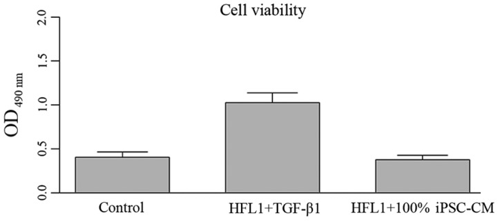
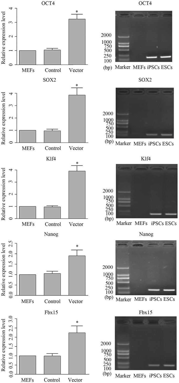
Therapeutic strategies based on stem cells have been shown to have potential in improving the condition of severe lung diseases. In this study, the suppressive effects of conditioned medium (CM) of induced pluripotent stem cells (iPSCs) on pulmonary fibroblast differentiation were investigated in a series of in vitro and in vivo experiments. Moreover, the underlying mechanisms through which iPSC-CM inhibited the differentiation of fibroblasts into myofibroblasts were explored as well. iPSCs were generated using a mouse 3-gene transfection method, myofibroblast-like cells were induced by incubating human fibroblasts with transforming growth factor-β1 (TGF-β1) and mouse models of pulmonary fibrosis (PF) were established by an injection of bleomycin. Based on these experiments, the effects of iPSC-CM on collagen accumulation, lung structure and the TGF-β1-mediated pathway were determined. It was found that treatment with iPSC-CM markedly reduced the proliferation of TGF-β1-exposed cells, and the activities of TGF-β1, Smad-2 and Smad-3. Accompanied by alterations in the expression of the indicated molecules, the lung structure of mice with PF was also markedly ameliorated. The present study confirmed the protective effects of iPSC-CM on lung tissue against PF, and it was also inferred that the ameliorating function of iPSC-CM on PF may be exerted through the blocking of TGF-β1/Smad signal transduction pathway.


Figures

Figure 1
Figure 1
Induced pluripotent stem cell-conditioned medium (iPSC-CM) had no impact on the viability of HFL1 cells. Control group, normal HFL1 cells. HFL1 + transforming growth factor-β1 (TGF-β1) group, HFL1 cells incubated with 5 ng/ml TGF-β1 for 24 h. HFL1 + 100% iPSC-CM, HLF1 cells incubated with 100% iPSC-CM for 24 h.
Figure 2
Figure 2
Induced pluripotent stem cell-conditioned medium (iPSC-CM) had no impact on the protein expression of proliferating cell nuclear antigen (PCNA), α-smooth muscle actin (α-SMA), collagen I, Smad-2, p-Smad-2, Smad-3 and p-Smad-3. Control group, normal HFL1 cells. HFL1 + transforming growth factor-β1 (TGF-β1) group, HFL1 cells incubated with 5 ng/ml TGF-β1 for 24 h. HFL1 + 100% iPSC-CM, HLF1 cells incubated with 100% iPSC-CM for 24 h.
Figure 3
Figure 3
Generation of induced pluripotent stem cells (iPSCs) using mouse 3-gene transfection. The bars represented the quantitative results of RT-qPCR of genes which were signature of mouse embryonic stem cells (ESCs). Images represent the expression patterns of the target genes compared with mouse embryonic fibroblasts (MEFs) and ESCs. *P<0.05, significantly different from MEFs.
Figure 4
Figure 4
In vitro osteogenic and adipogenic differentiation of induced pluripotent stem cells (iPSCs) induced by mouse 3-gene transfection. (A) Representative images of osteogenic differentiation of iPSCs as detected by Alizarin Red S staining; osteogenic iPSCs were stained red. (B) Representative images of adipogenic differentiation of iPSCs as detected by Oil Red O staining; adipogenic iPSCs were stained red. Magnification, ×400.
Figure 5
Figure 5
Administration of induced pluripotent stem cell-conditioned medium (iPSC-CM) inhibits the transforming growth factor-β1 (TGF-β1) induced proliferation of HLF1 cells. (A) MTT assay for cell viability; quantitative results are shown. (B) Representative blots and quantitative results of western blot analysis of proliferating cell nuclear antigen (PCNA). aP<0.05, significantly different from the control group; bP<0.05, significantly different from the TGF-β1 group; cP<0.05, significantly different from the 30% iPSC-CM group.
Figure 6
Figure 6
Administration of induced pluripotent stem cell-conditioned medium (iPSC-CM) inhibits the transforming growth factor-β1 (TGF-β1)-induced differentiation of HFL1 cells into myofibroblasts. (A) Representative blots and quantitative results of western blot analysis of collagen I. (B) Quantitative results of RT-qPCR of collagen I. (C) Representative blots and quantitative results of western blot analysis of α-smooth muscle actin (α-SMA). (D) Quantitative results of RT-qPCR of α-SMA. aP<0.05, significantly different from the control group; bP<0.05, significantly different from the TGF-β1 group; cP<0.05, significantly different from the 30% iPSC-CM group; dP<0.05, significantly different from the 50% iPSC-CM group.
Figure 7
Figure 7
Induced pluripotent stem cell-conditioned medium (iPSC-CM) exerts an inhibitory effect on transforming growth factor-β1 (TGF-β1)-induced processes via blocking the Smad signal transduction pathway in vitro. (A) Representative blots and quantitative results of western blot analysis of p-Smad-2 and Smad-2. (B) Representative blots and quantitative results of western blot analysis of p-Smad-3 and Smad-3. aP<0.05, significantly different from the control group; bP<0.05, significantly different from the TGF-β1 group; cP<0.05, significantly different from the 30% iPSC-CM group; dP<0.05, significantly different from the 50% iPSC-CM group.
Figure 8
Figure 8
Administration of induced pluripotent stem cell-conditioned medium (iPSC-CM) attenuates injury to lung tissues due to pulmonary fibrosis (PF) and reduces collagen accumulation. (A) Representative image of H&E staining (×200 magnification); the nuclei in myocardial tissue were stained blue and cytoplasma were stained red. Green arrow indicates patched areas and yellow arrow indicates neutrophilic alveolitis. (B) Statistical analysis results of ELISA of collagen I. (C) Representative image of Masson's staining, collagen I was stained blue. (D) Statistical analysis results of the detection of hydroxyproline. (E) Representative blots and quantitative results of western blot analysis of α-smooth muscle actin (α-SMA) in model mice. aP<0.05, significantly different from the control group; bP<0.05, significantly different from the PF group.
Figure 9
Figure 9
Administration of induced pluripotent stem cell-conditioned medium (iPSC-CM) reduces the expression of transforming growth factor-β1 (TGF-β1) in model mice. (A) Statistical analysis results of ELISA results of TGF-β1 production in BALF. (B) Representative blots and quantitative results of western blot analysis of TGF-β1 expression in lung tissue. (C) Quantitative results of RT-qPCR of TGF-β1 expression in lung tissue. aP<0.05, significantly different from the control group; bP<0.05, significantly different from the pulmonary fibrosis (PF) group.
Figure 10
Figure 10
Induced pluripotent stem cell-conditioned medium (iPSC-CM) exerts an inhibitory effect on transforming growth factor-β1 (TGF-β1)-induced processes via blocking the Smad signal transduction pathway in vivo. (A) Representative blots and quantitative results of western blot analysis of p-Smad-2 and Smad-2 expression in lung tissue. (B) Representative blots and quantitative results of western blot analysis of p-Smad-3 and Smad-3 expression in lung tissue. aP<0.05, significantly different from the control group; bP<0.05, significantly different from the pulmonary fibrosis (PF) group.

References

    1. Ware LB, Matthay MA. The acute respiratory distress syndrome. N Engl J Med. 2000;342:1334–1349. doi: 10.1056/NEJM200005043421806. - DOI - PubMed
    1. Perez A, Rogers RM, Dauber JH. The prognosis of idiopathic pulmonary fibrosis. Am J Respir Cell Mol Biol. 2003;29(Suppl 3):S19–S26. - PubMed
    1. Phan SH. The myofibroblast in pulmonary fibrosis. Chest. 2002;122(Suppl 6):286S–289S. doi: 10.1378/chest.122.6_suppl.286S. - DOI - PubMed
    1. Evans RA, Tian YC, Steadman R, Phillips AO. TGF-β1-mediated fibroblast-myofibroblast terminal differentiation-the role of Smad proteins. Exp Cell Res. 2003;282:90–100. doi: 10.1016/S0014-4827(02)00015-0. - DOI - PubMed
    1. Sime PJ, Xing Z, Graham FL, Csaky KG, Gauldie J. Adeno-vector-mediated gene transfer of active transforming growth factor-beta1 induces prolonged severe fibrosis in rat lung. J Clin Invest. 1997;100:768–776. doi: 10.1172/JCI119590. - DOI - PMC - PubMed

MeSH terms