. 2018 Jan;41(1):33-42.
doi: 10.3892/ijmm.2017.3200. Epub 2017 Oct 19.

Atorvastatin protects the proliferative ability of human umbilical vein endothelial cells inhibited by angiotensin II by changing mitochondrial energy metabolism

Affiliations

Atorvastatin protects the proliferative ability of human umbilical vein endothelial cells inhibited by angiotensin II by changing mitochondrial energy metabolism

Ye Chang et al. Int J Mol Med. 2018 Jan.

Abstract

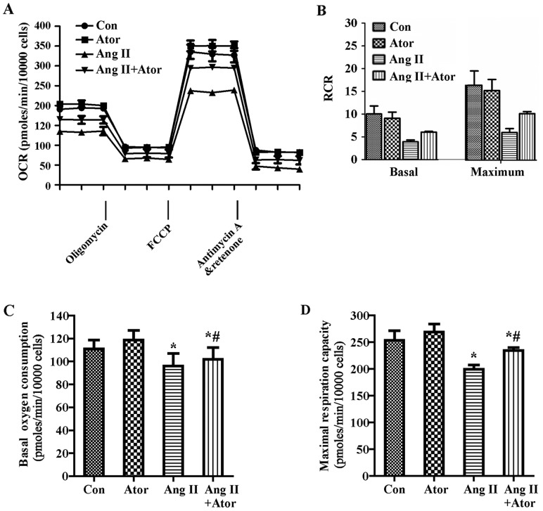
This study aimed to explore whether angiotensin II (Ang II) inhibits the proliferation of human umbilical vein endothelial cells (HUVECs) by changing mitochondrial energy metabolism, and whether atorvastatin has a protective role via restoration of endothelial function. HUVECs were treated with 1 µM Ang II alone or with 10 µM atorvastatin for 24 h. Proliferation was detected by MTT assay, cell counting, 5‑ethynyl‑2'‑deoxyuridine assay and real‑time cell analyzer. Mitochondrial energy metabolism including oxygen consumption rate and extracellular acidification rate were measured using a Seahorse metabolic flux analyzer. Mitochondrial membrane potential was detected under fluorescence microscope following staining with tetramethylrhodamine. Respiratory chain complexes I‑V were detected using western blotting. The current study showed that Ang II inhibits the proliferation of HUVECs. Results from the Seahorse metabolic flux analyzer indicated that Ang II decreased basal oxygen consumption, maximal respiration capacity, spare respiration capacity, adenosine triphosphate‑linked respiration and non‑mitochondrial respiration. By contrast, Ang II increased the proton leak. Additionally, Ang II increased glycolysis, glycolytic capacity and non‑glycolytic acidification. Furthermore, these effects were all suppressed by atorvastatin. The results indicated that atorvastatin prevents cellular energy metabolism switching from oxidative phosphorylation to glycolysis induced by Ang II and protected the proliferative ability of HUVECs.


Figures

Figure 1
Figure 1
Atorvastatin attenuated Ang II-inhibited proliferation in human umbilical vein endothelial cells. Cells were cultured in the presence of 1 µM Ang II and/or 10 µM atorvastatin. (A) Cell viability was detected by MTT assay. (B) Cells were plated on 6-well plate and cell numbers were counted daily for 4 days. (C) Cell proliferation was detected by EdU assay and the red staining represents the new cells and (D) quantified by EdU incorporation (%). (E) Cell index was measured by real-time cell analyzer. Results are presented as the mean ± standard deviation of at least three experiments. *P<0.05 vs. control; #P<0.05 vs. Ang II. Con, control; Ator, atorvastatin; Ang II, angiotensin II; EdU, 5-ethynyl-2′-deoxyuridine.
Figure 2
Figure 2
Measurement of mitochondrial function in HUVECs. The experiments were performed using the XF24 extracellular flux analyzer, and the flow chart showed the measurement of (A) OCR and (B) ECAR. The optimal number (10,000, 20,000, 40,000 and 80,000 cells/well) of HUVECs were seeded in the Seahorse Bioscience microplates. (C) OCR and (D) ECAR were measured and plotted as a function of cell seeding number. Results are presented as the mean ± standard deviation (n=4). HUVECs, human umbilical vein endothelial cells; OCR, oxygen consumption rate; FCCP, carbonyl cyanide-4-(trifluoromethoxy)phenylhydrazone; ECAR, extracellular acidification rate; 2-DG, 2-deoxy-D-glucose.
Figure 3
Figure 3
Effect of Ang II and/or atorvastatin on mitochondrial aerobic metabolism in HUVECs. HUVECs were seeded in the Seahorse Bioscience microplates (10,000 cells/well). (A) The OCR was conducted using Mito Stress Test kit. After adherence for 4 h, 1 µM Ang II and/or 10 µM atorvastatin was added into the microplates for co-incubation with cells for 24 h with subsequent injection of 1 µM oligomycin, 0.5 µM FCCP and 0.5 µM rotenone and antimycin A. (B) The contribution of associated parameters including RCR, (C) basal oxygen consumption, (D) maximal oxygen consumption, (E) spare respiration capacity, (F) ATP-linked oxygen consumption, (G) proton leak and (H) non-mitochondrial oxygen consumption to the total cellular oxygen consumption was plotted, respectively. Each data point represented an OCR measurement. Results shown represent mean ± standard deviation (n=4). *P<0.05 vs. control; #P<0.05 vs. Ang II. HUVECs, human umbilical vein endothelial cells; Con, control; Ang II, angiotensin II; Ator, atorvastatin; OCR, oxygen consumption rate; RCR, respiratory control ratio.
Figure 3
Figure 3
Effect of Ang II and/or atorvastatin on mitochondrial aerobic metabolism in HUVECs. HUVECs were seeded in the Seahorse Bioscience microplates (10,000 cells/well). (A) The OCR was conducted using Mito Stress Test kit. After adherence for 4 h, 1 µM Ang II and/or 10 µM atorvastatin was added into the microplates for co-incubation with cells for 24 h with subsequent injection of 1 µM oligomycin, 0.5 µM FCCP and 0.5 µM rotenone and antimycin A. (B) The contribution of associated parameters including RCR, (C) basal oxygen consumption, (D) maximal oxygen consumption, (E) spare respiration capacity, (F) ATP-linked oxygen consumption, (G) proton leak and (H) non-mitochondrial oxygen consumption to the total cellular oxygen consumption was plotted, respectively. Each data point represented an OCR measurement. Results shown represent mean ± standard deviation (n=4). *P<0.05 vs. control; #P<0.05 vs. Ang II. HUVECs, human umbilical vein endothelial cells; Con, control; Ang II, angiotensin II; Ator, atorvastatin; OCR, oxygen consumption rate; RCR, respiratory control ratio.
Figure 4
Figure 4
Effect of Ang II and/or atorvastatin on glycolytic function in HUVECs. HUVECs were seeded in the Seahorse Bioscience microplates (10,000 cells/well). (A) The ECAR was conducted using XF Glycolysis Stress Test kit. After adherence for 4 h, 1 µM Ang II and/or 10 µM atorvastatin was added into the microplates for co-incubation with cells for 24 h with subsequent injection of 15 mM glucose, 2 µM oligomycin and 50 mM 2-DG. (B) The contribution of associated parameters including non-glycolytic acidification, (C) glycolysis, (D) glycolytic capacity and (E) glycolytic reserve to the total ECAR was plotted, respectively. Each data point represented an ECAR measurement. Results shown represent mean ± standard deviation (n=4). *P<0.05 vs. control; #P<0.05 vs. Ang II. HUVECs, human umbilical vein endothelial cells; ECAR, extracellular acidification rate; Con, control; Ang II, angiotensin II; Ator, atorvastatin; 2-DG, 2-deoxy-D-glucose.
Figure 5
Figure 5
Effect of Ang II and/or atorvastatin on mitochondrial membrane potential and mitochondrial respiration chain complexes in HUVECs. (A) The mitochondria of the HUVECs were marked with TMRE (red), and the HUVECs nuclei were stained with Hoechst 33342 (blue). (B) Mitochondrial respiration chain complexes I–V were detected by western blotting and GAPDH served as a control, and (C) the band intensity was quantified using ImageJ 1.47 software, normalized by the control. Results are presented as the mean ± standard deviation for at least three experiments. *P<0.05 vs. control; #P<0.05 vs. Ang II. HUVECs, human umbilical vein endothelial cells; TMRE, tetramethylrhodamine; Con, control; Ator, atorvastatin; Ang II, angiotensin II.

Similar articles

Cited by

References

    1. Whaley-Connell A, Johnson MS, Sowers JR. Aldosterone: role in the cardiometabolic syndrome and resistant hypertension. Prog Cardiovasc Dis. 2010;52:401–409. doi: 10.1016/j.pcad.2009.12.004. - DOI - PMC - PubMed
    1. Aroor AR, Demarco VG, Jia G, Sun Z, Nistala R, Meininger GA, Sowers JR. Front Endocrinol. Vol. 4. Lausanne: 2013. The role of tissue renin-angiotensin-aldosterone system in the development of endothelial dysfunction and arterial stiffness; p. 161. - DOI - PMC - PubMed
    1. Schiffrin EL, Touyz RM. From bedside to bench to bedside: role of renin-angiotensin-aldosterone system in remodeling of resistance arteries in hypertension. Am J Physiol Heart Circ Physiol. 2004;287:H435–H446. doi: 10.1152/ajpheart.00262.2004. - DOI - PubMed
    1. Touyz RM. The role of angiotensin II in regulating vascular structural and functional changes in hypertension. Curr Hypertens Rep. 2003;5:155–164. doi: 10.1007/s11906-003-0073-2. - DOI - PubMed
    1. Stoll M, Steckelings UM, Paul M, Bottari SP, Metzger R, Unger T. The angiotensin AT2-receptor mediates inhibition of cell proliferation in coronary endothelial cells. J Clin Invest. 1995;95:651–657. doi: 10.1172/JCI117710. - DOI - PMC - PubMed

MeSH terms