. 2018 Jan;17(1):898-910.
doi: 10.3892/mmr.2017.7998. Epub 2017 Nov 7.

Roles and mechanisms of TRPC3 and the PLCγ/PKC/CPI-17 signaling pathway in regulating parturition

Affiliations

Roles and mechanisms of TRPC3 and the PLCγ/PKC/CPI-17 signaling pathway in regulating parturition

Jing Chen et al. Mol Med Rep. 2018 Jan.

Erratum in

Abstract

The aim of the current study was to investigate the role of phospholipase C (PLC)γ/protein kinase C (PKC)/C‑kinase‑activated protein phosphatase‑1 (CPI‑17) signaling pathways in uterine smooth muscle during parturition. Samples of uterine tissue were collected from pregnant patients who underwent a caesarean section for preterm delivery, full‑term delivery with labor onset, full‑term delivery without labor onset, and from a non‑pregnant control group undergoing surgery for cervical intraepithelial neoplasia III. Immunohistochemistry, and western blotting were used to assess the association between TRPC3 levels and parturition and the influence of calcium ion channels. In addition, pregnant mice were used to explore the effect of uterine canonical transient receptor potential 3 (TRPC3) expression on the parturition‑triggering mechanism and PLCγ/PKC/CPI‑17 signaling pathways. Pregnant mouse uterine smooth muscle cells were cultivated, with and without TRPC3 silencing, and the expression levels of PLCγ, PKC and CPI‑17, the upstream and downstream factors of the TRPC3 pathway, were measured in pregnant mouse uterine smooth muscle cells, in order to provide a theoretical basis for the prevention and treatment of premature labor. In the preterm and full‑term without labor onset patient groups, the TRPC3 gene expression in the mSMCs was significantly overexpressed when compared with the non‑pregnant group (P<0.05); however, TRPC3 expression was not elevated in the full‑term with labor onset group, exhibiting no significant difference compared with the non‑pregnant group (P>0.05). During pregnancy, compared with the non‑pregnant controls, Cav1.2, Cav3.1 and Cav3.2 gene expression levels were markedly increased (P<0.05) in mSMCs from the preterm delivery group and the full‑term with labor onset group, however were non‑significantly increased in the full‑term without labor onset group. The level of TRPC3 was highest in the preterm group, while the levels of Cav1.2, Cav3.1 and Cav3.2 were highest in the full‑term with labor onset group. In the preterm, LPS‑treated preterm and full‑term groups, TRPC3, MAPK, ERK1/2, P‑ERK, Cav3.2, Cav3.1 and Cav1.2 were all expressed at higher levels than in the unfertilized group. In the LPS‑treated preterm group, the levels of TRPC3, MAPK, ERK1/2, P‑ERK, Cav3.2, Cav3.1 and Cav1.2 were increased compared with the preterm group. Furthermore, following transfection of small interfering TRPC3 (siTRPC3) into cells, it was demonstrated that the levels of TRPC3, PLCγ, PKC, CPI‑17, P‑CPI‑17, Cav1.2, Cav3.1 and Cav3.2 expression were lower in the LPS siTRPC3 group when compared with that of the LPS‑treated untransfected control group.


Figures

Figure 1.
Figure 1.
Expression levels of TRPC3, Cav1.2, Cav.3.1 and Cav3.2 in human myometrial smooth muscle cells derived from the non-pregnant, full-term without labor onset, preterm, and full-term with labor onset patient groups (n=20/group). (A) Western blot analysis of protein expression levels in the different groups. GAPDH was used as the loading control. Quantified western blot analyses of relative expression levels of (B) TRPC3, (C) Cav1.2, (D) Cav.3.1 and (E) Cav3.2. Data are presented as the mean ± standard error. *P<0.05 vs. non-pregnant group. TRPC3, canonical transient receptor potential 3.
Figure 2.
Figure 2.
Expression levels of TRPC3, MAPK, P-ERK, Cav1.2, Cav.3.1 and Cav3.2 in mouse myometrial smooth muscle cells derived from the non-pregnant, preterm, infected preterm and full-term groups (n=10/group). (A) Western blot analysis of protein expression levels in the different groups. GAPDH was used as the loading control. Quantified protein expression levels of (B) TRPC3, (C) MAPK, (D) P-ERK, (E) Cav1.2, (F) Cav.3.1 and (G) Cav3.2. Data are presented as the mean ± standard error. *P<0.05 vs. non-pregnant group. TRPC3, canonical transient receptor potential 3; MAPK, mitogen-activated protein kinase; ERK, extracellular signal-related kinase; P-, phosphorylated.
Figure 3.
Figure 3.
Immunostaining of smooth muscle actin in mouse uterine smooth muscle cells, and transfection with TRPC3 siRNA. (A) Mouse uterine smooth muscle cells. (B) Immunostaining of TRPC3 in mouse uterine smooth muscle cells. (C) The expression of TRPC3 detected by western blotting. TRPC3, canonical transient receptor potential 3; siRNA, small interfering RNA.
Figure 4.
Figure 4.
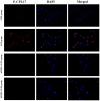
Expression of TRPC3 protein with and without TRPC3-siRNA transfection of murine uterine smooth muscle cells. CON, untransfected non-infected with LPS; LPS, infected with LPS; siTRPC3 CON, transfected with siTRPC3 and non-infected with LPS; siTRPC3 LPS, transfected with siTRPC3 and infected with LPS. TRPC3, canonical transient receptor potential 3; siRNA, small interfering RNA; LPS, lipopolysaccharides; CON, control.
Figure 5.
Figure 5.
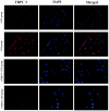
Expression of PLCγ protein with and without TRPC3-siRNA transfection in murine uterine smooth muscle cells. CON, untransfected non-infected with LPS; LPS, infected with LPS; siTRPC3 CON, transfected with siTRPC3 and non-infected with LPS; siTRPC3 LPS, transfected with siTRPC3 and infected with LPS. PLC, phospholipase C; TRPC3, canonical transient receptor potential 3; siRNA, small interfering RNA; LPS, lipopolysaccharides; CON, control.
Figure 6.
Figure 6.
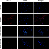
Expression of PKC protein with and without TRPC3-siRNA transfection in murine uterine smooth muscle cells. CON, untransfected non-infected with LPS; LPS, infected with LPS; siTRPC3 CON, transfected with siTRPC3 and non-infected with LPS; siTRPC3 LPS, transfected with siTRPC3 and infected with LPS. PKC, protein kinase C; TRPC3, canonical transient receptor potential 3; siRNA, small interfering RNA; LPS, lipopolysaccharides; CON, control.
Figure 7.
Figure 7.
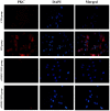
Expression of CPI-17 protein with and without TRPC3-siRNA transfection in murine uterine smooth muscle cells. CON, untransfected non-infected with LPS; LPS, infected with LPS; siTRPC3 CON, transfected with siTRPC3 and non-infected with LPS; siTRPC3 LPS, transfected with siTRPC3 and infected with LPS. CPI-17, C-kinase-activated protein phosphatase-1 inhibitor; TRPC3, canonical transient receptor potential 3; siRNA, small interfering RNA; LPS, lipopolysaccharides; CON, control.
Figure 8.
Figure 8.
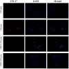
Expression of P-CPI-17 protein with and without TRPC3-siRNA transfection murine uterine smooth muscle cells. CON, untransfected non-infected with LPS; LPS, infected with LPS; siTRPC3 CON, transfected with siTRPC3 and non-infected with LPS; siTRPC3 LPS, transfected with siTRPC3 and infected with LPS. P-, phosphorylated; CPI-17, C-kinase-activated protein phosphatase-1 inhibitor; TRPC3, canonical transient receptor potential 3; siRNA, small interfering RNA; LPS, lipopolysaccharides; CON, control.
Figure 9.
Figure 9.
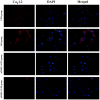
Expression of Cav1.2 protein with and without TRPC3-siRNA transfection in murine uterine smooth muscle cells. CON, untransfected non-infected with LPS; LPS, infected with LPS; siTRPC3 CON, transfected with siTRPC3 and non-infected with LPS; siTRPC3 LPS, transfected with siTRPC3 and infected with LPS. TRPC3, canonical transient receptor potential 3; siRNA, small interfering RNA; LPS, lipopolysaccharides; CON, control.
Figure 10.
Figure 10.
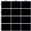
Expression of Cav3.1 protein with and without TRPC3-siRNA transfection in murine uterine smooth muscle cells. CON, untransfected non-infected with LPS; LPS, infected with LPS; siTRPC3 CON, transfected with siTRPC3 and non-infected with LPS; siTRPC3 LPS, transfected with siTRPC3 and infected with LPS. TRPC3, canonical transient receptor potential 3; siRNA, small interfering RNA; LPS, lipopolysaccharides; CON, control.
Figure 11.
Figure 11.
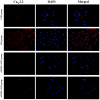
Expression of Cav3.2 protein with and without TRPC3-siRNA transfection in murine uterine smooth muscle cells. CON, untransfected non-infected with LPS; LPS, infected with LPS; siTRPC3 CON, transfected with siTRPC3 and non-infected with LPS; siTRPC3 LPS, transfected with siTRPC3 and infected with LPS. TRPC3, canonical transient receptor potential 3; siRNA, small interfering RNA; LPS, lipopolysaccharides; CON, control.
Figure 12.
Figure 12.
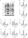
(A) Western blot analysis of proteins in mouse myometrial smooth muscle cells with and without TRPC3-siRNA transfection. GAPDH was used as the loading control. Quantified protein expression levels of (B) TRPC3, (C) PLCγ, (D) PKC, (E) CPI-17, (F) P-CPI-17, (G) Cav1.2, (H) Cav3.1 and (I) Cav3.2. Data are presented as the mean ± standard error. *P<0.05 vs. CON group. CON, untransfected non-infected with LPS; LPS, infected with LPS; siTRPC3 CON, transfected with siTRPC3 and non-infected with LPS; siTRPC3 LPS, transfected with siTRPC3 and infected with LPS. TRPC3, canonical transient receptor potential 3; siRNA, small interfering RNA; PLC, phospholipase C; PKC, protein kinase C; CPI-17, C-kinase-activated protein phosphatase-1 inhibitor; LPS, lipopolysaccharides; CON, control.

References

    1. Senturk MB, Cakmak Y, Gündoğdu M, Polat M, Atac H. Does performing cesarean section after onset of labor has positive effect on neonatal respiratory disorders? J Matern Fetal Neonatal Med. 2016;29:2457–2460. - PubMed
    1. Hanley GE, Munro S, Greyson D, Gross MM, Hundley V, Spiby H, Janssen PA. Diagnosing onset of labor: A systematic review of definitions in the research literature. BMC Pregnancy Childbirth. 2016;16:71. doi: 10.1186/s12884-016-0857-4. - DOI - PMC - PubMed
    1. Ohno Y, Terauchi M, Tamakoshi K, Shiozaki A, Saito S. The risk factors for labor onset hypertension. Hypertens Res. 2016;39:260–265. doi: 10.1038/hr.2015.112. - DOI - PubMed
    1. Neal JL, Lowe NK. Physiologic partograph to improve birth safety and outcomes among low-risk, nulliparous women with spontaneous labor onset. Med Hypotheses. 2012;78:319–326. doi: 10.1016/j.mehy.2011.11.012. - DOI - PMC - PubMed
    1. Kiesewetter B, Lehner R. Maternal outcome monitoring: Induction of labor versus spontaneous onset of labor-a retrospective data analysis. Arch Gynecol Obstet. 2012;286:37–41. doi: 10.1007/s00404-012-2239-0. - DOI - PubMed