Comparative Study
. 2017 Nov 7;17(1):728.
doi: 10.1186/s12885-017-3751-1.

MicroRNA expression patterns in canine mammary cancer show significant differences between metastatic and non-metastatic tumours

Affiliations
Comparative Study

MicroRNA expression patterns in canine mammary cancer show significant differences between metastatic and non-metastatic tumours

Malgorzata Bulkowska et al. BMC Cancer. .

Abstract

Background: MicroRNAs may act as oncogenes or tumour suppressor genes, which make these small molecules potential diagnostic/prognostic factors and targets for anticancer therapies. Several common oncogenic microRNAs have been found for canine mammary cancer and human breast cancer. On account of this, large-scale profiling of microRNA expression in canine mammary cancer seems to be important for both dogs and humans.

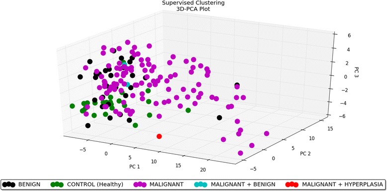
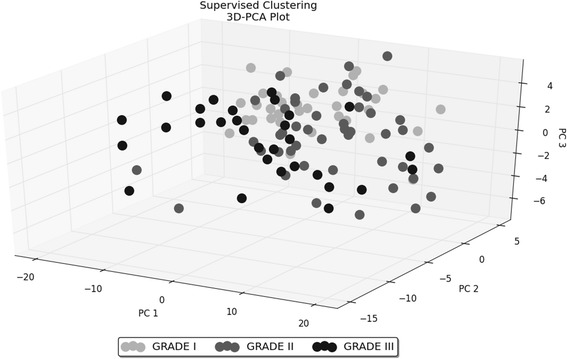
Methods: Expression profiles of 317 microRNAs in 146 canine mammary tumours of different histological type, malignancy grade and clinical history (presence/absence of metastases) and in 25 control samples were evaluated. The profiling was performed using microarrays. Significance Analysis of Microarrays test was applied in the analysis of microarray data (both unsupervised and supervised data analyses were performed). Validation of the obtained results was performed using real-time qPCR. Subsequently, predicted targets for the microRNAs were searched for in miRBase.

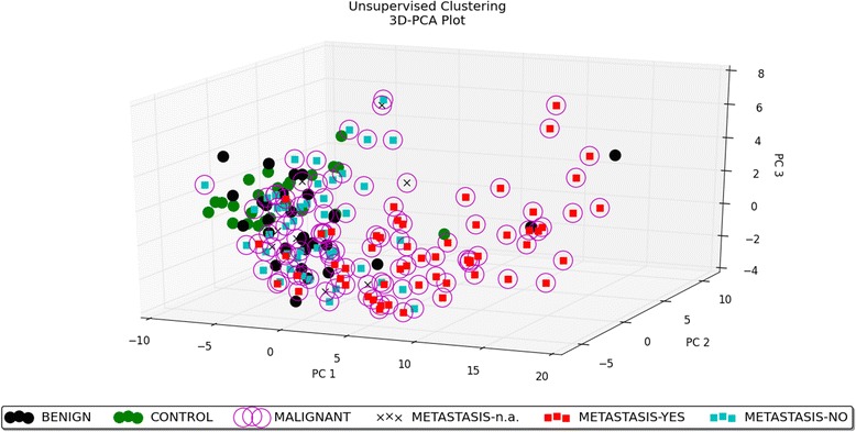
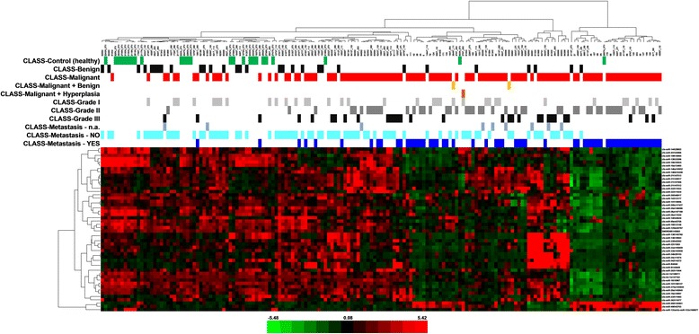
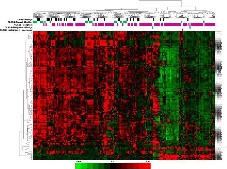
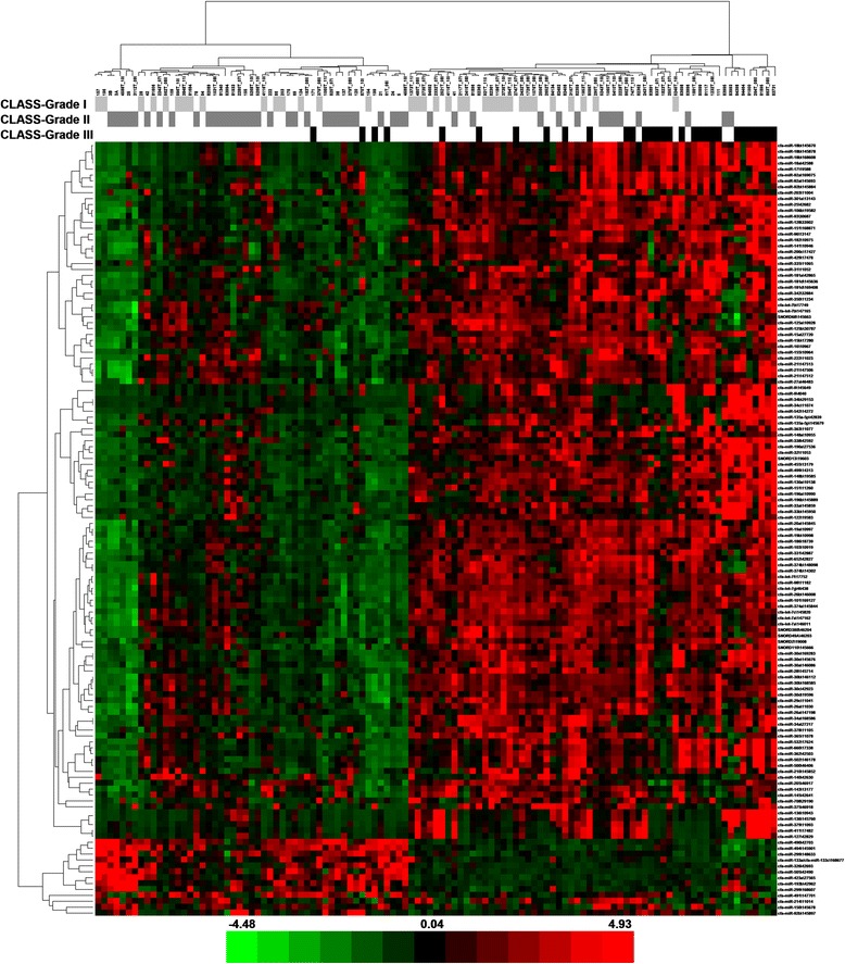
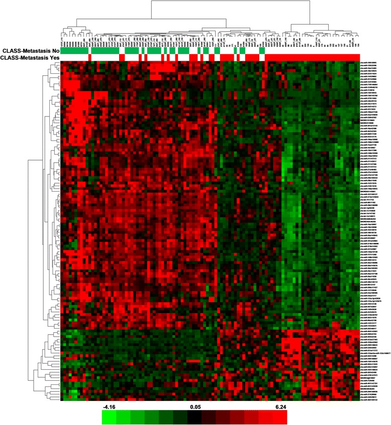
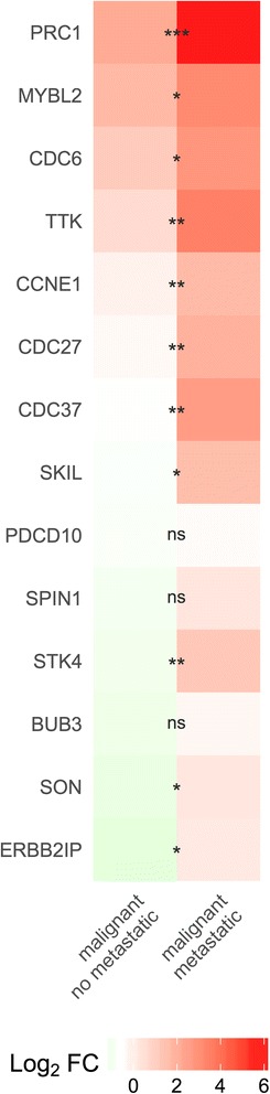
Results: Results of the unsupervised analysis indicate that the primary factor separating the samples is the metastasis status. Predicted targets for microRNAs differentially expressed in the metastatic vs. non-metastatic group are mostly engaged in cell cycle regulation, cell differentiation and DNA-damage repair. On the other hand, the supervised analysis reveals clusters of differentially expressed microRNAs unique for the tumour type, malignancy grade and metastasis factor.

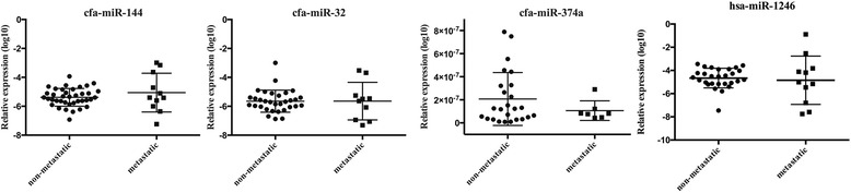
Conclusions: The most significant difference in microRNA expression was observed between the metastatic and non-metastatic group, which suggests a more important role of microRNAs in the metastasis process than in the malignant transformation. Moreover, the differentially expressed microRNAs constitute potential metastasis markers. However, validation of cfa-miR-144, cfa-miR-32 and cfa-miR-374a levels in blood samples did not follow changes observed in the non-metastatic and metastatic tumours.

Keywords: Canine mammary cancer; Human breast cancer; microRNA.


Conflict of interest statement

Ethics approval and consent to participate

Poland: The dogs’ owners in Poland gave oral permission for the use of their animals’ tissue and blood samples for this work. Tissue sampling in Poland was approved by the III Local Ethical Committee (approval No. 8/2012, 17.01.2012) of the Warsaw University of Life Sciences. Blood samples in Poland were collected according to the Polish legal act concerning experiments performed on animals (Ustawa o doświadczeniach na zwierzętach z‬ dnia 21 stycznia 2005 r. (Dz. U. z 2005 r. Nr 33, poz.‬ 289 z późn.zm.)), no written approval of the study by the Local Ethical Committee was required. Sweden: The dogs’ owners in Sweden signed a written agreement for a post-mortem examination of the dogs and sampling of the tumours for scientific purposes. In Sweden, there were no regulations concerning ethical approval of experiments performed on animals at the time of the tumour sampling (1985–1987). Germany: The dogs’ owners in Germany gave oral permission for the use of their animals’ tissue samples for scientific purposes. Tissue sampling in Germany was performed with the approval of animal welfare authorities of the Veterinary College of the Freie Universitaet Berlin. Surgical excision of tumour biopsies was part of the tumour treatment according to the state of the art treatment and solely to improve the animals’ welfare. Furthermore, the animals were under full anaesthesia and not exposed to any additional manipulation due to the inclusion in this study. Therefore, no written approval of the sampling by any ethics committee was required.

Consent for publication

Not applicable.

Competing interests

The authors declare that they have no competing interests.

Publisher’s Note

Springer Nature remains neutral with regard to jurisdictional claims in published maps and institutional affiliations.

Figures

Fig. 1
Fig. 1
3D–PCA plot − unsupervised analysis. PCA performed on all the samples and on the top 50 microRNAs with the highest standard deviation. The normalized log-transformed Hy3 values were used for the analysis. The features were shifted to be zero centred, (i.e. the mean value across samples was shifted to 0) and scaled to have unit variance (i.e. the variance across samples was scaled to 1) before the analysis. PCA plot reveals the distinct sample clusters for metastatic tumours, non-metastatic tumours and the control group
Fig. 2
Fig. 2
Heat map − unsupervised hierarchical clustering. Clustering performed on all the samples and on the top 50 microRNAs with the highest standard deviation. The normalized log-transformed Hy3 values were used for the analysis. The colour scale illustrates the relative expression level of a microRNA across all samples: green colour represents an expression level below the mean and red colour represents an expression level above the mean. Legend: n.a. − data not available
Fig. 3
Fig. 3
Heat map − tumour type. The scaled expression of the differentially expressed microRNAs for 171 samples and the relationship among the samples in terms of microRNAs found to be differentially expressed for the tumour type factor; Significance Analysis of Microarrays (SAM) test; the colour scale illustrates the relative expression level of a microRNA across all samples: green colour represents an expression level below the mean and red colour represents an expression level above the mean. The most distinct differences in microRNA profile are between the control and malignant group − microRNAs mostly down-regulated in the malignant group
Fig. 4
Fig. 4
Heat map − grade of malignancy. The scaled expression of the differentially expressed microRNAs for 111 samples and the relationship among the samples in terms of microRNAs found to be differentially expressed for the malignancy grade factor; Significance Analysis of Microarrays (SAM) test; the colour scale illustrates the relative expression level of a microRNA across all samples: green colour represents an expression level below the mean and red colour represents an expression level above the mean. MicroRNAs are mostly up-regulated in the grade III group in comparison with the grade II group
Fig. 5
Fig. 5
Heat map − metastasis. The scaled expression of the differentially expressed microRNAs for 107 samples and the relationship among the samples in terms of microRNAs found to be differentially expressed for the metastasis factor; Significance Analysis of Microarrays (SAM) test; the colour scale illustrates the relative expression level of a microRNA across all samples: green colour represents an expression level below the mean and red colour represents an expression level above the mean. MicroRNAs are mostly down-regulated in the metastatic group
Fig. 6
Fig. 6
3D–PCA plot − tumour type. PCA plot performed on 171 samples and 123 differentially expressed microRNAs for the tumour type factor; Significance Analysis of Microarrays (SAM) test. PCA plot reveals the distinct sample clusters for malignant tumours, benign tumours and the control group
Fig. 7
Fig. 7
3D–PCA plot − grade of malignancy. PCA plot performed on 111 samples and 131 differentially expressed microRNAs for the malignancy grade factor; Significance Analysis of Microarrays (SAM) test
Fig. 8
Fig. 8
3D–PCA plot − metastasis. PCA plot performed on 107 samples and 124 differentially expressed microRNAs for the metastasis factor; Significance Analysis of Microarrays (SAM) test. PCA plot reveals the distinct sample clusters for metastatic tumours and non-metastatic tumours
Fig. 9
Fig. 9
Heat map − selected targets for microRNAs deregulated in metastatic canine mammary cancer. The scaled expression of CDC6, CCNE1, MYBL2, PDCD10, ERBB2IP, SON, STK4, CDC27, PRC1, CDC37, TTK, SKIL, BUB3 and SPIN1. The analysis performed on ten malignant non-metastatic tumours and ten malignant metastatic tumours. Statistical analysis was made on Ct values normalized with a housekeeping gene; one-way ANOVA followed by Tukey’s HSD post hoc tests. Legend: log2 FC – log base 2 from fold change, * – p value <0.05, ** – p value <0.01, *** – p value <0.001
Fig. 10
Fig. 10
Expression of selected microRNAs in plasma samples from dogs with non-metastatic and metastatic tumours. Relative expression of cfa-miR-144, cfa-miR-32 cfa-miR-374a and hsa-miR-1246 in plasma samples from dogs with non-metastatic and metastatic tumours. The statistical analysis was performed using Prism version 5.00 software (GraphPad Software, USA). An unpaired, non-parametric Mann-Whitney test was applied. Values are mean ± SD

References

    1. Egenvall A, Bonnett BN, Ohagen P, Olson P, Hedhammar A, von Euler H. Incidence of and survival after mammary tumors in a population of over 80,000 insured female dogs in Sweden from 1995 to 2002. Prev Vet Med. 2005;69:109–127. doi: 10.1016/j.prevetmed.2005.01.014. - DOI - PubMed
    1. Misdorp W. In: Tumors of the mammary gland. 4. Meuten DJ, editor. Ames: Iowa State Press; 2002. pp. 575–606.
    1. Shafiee R, Javanbakht J, Atyabi N, Kheradmand P, Kheradmand D, Bahrami A, et al. Diagnosis, classification and grading of canine mammary tumours as a model to study human breast cancer: an Clinico-Cytohistopathological study with environmental factors influencing public health and medicine. Cancer Cell Int. 2013;13:79. doi: 10.1186/1475-2867-13-79. - DOI - PMC - PubMed
    1. Weigelt B, Horlings HM, Kreike B, Hayes MM, Hauptmann M, Wessels LF, et al. Refinement of breast cancer classification by molecular characterization of histological special types. J Pathol. 2008;216:141–150. doi: 10.1002/path.2407. - DOI - PubMed
    1. Gilbertson SR, Kurzman ID, Zachrau RE, Hurvitz AI, Black MM. Canine mammary epithelial neoplasms: biologic implications of morphologic characteristics assessed in 232 dogs. Vet Pathol. 1983;20:127–142. doi: 10.1177/030098588302000201. - DOI - PubMed

Publication types

MeSH terms