. 2017 Nov 7;36(1):156.
doi: 10.1186/s13046-017-0623-0.

STAT3-blocked whole-cell hepatoma vaccine induces cellular and humoral immune response against HCC

Affiliations

STAT3-blocked whole-cell hepatoma vaccine induces cellular and humoral immune response against HCC

Qiuju Han et al. J Exp Clin Cancer Res. .

Abstract

Background: Whole-cell tumor vaccines have shown much promise; however, only limited success has been achieved for the goal of eliciting robust tumor-specific T-cell responses.

Methods: Hepatocellular carcinoma (HCC) cells, H22 and Hepa1-6, were modified by blocking the STAT3 signaling pathway with a STAT3 decoy oligodeoxynucleotide, and the immunogenicity and possibility of using these cell lysates as a vaccine were evaluated.

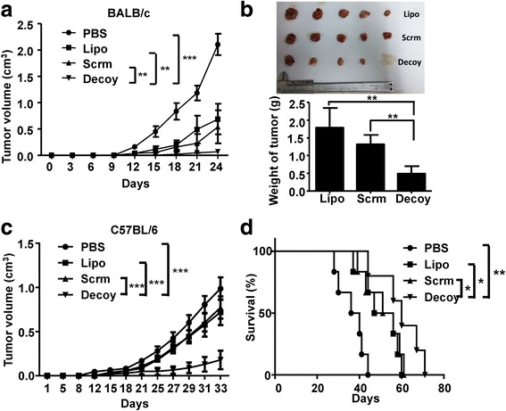
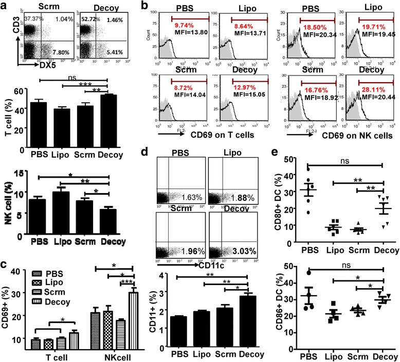
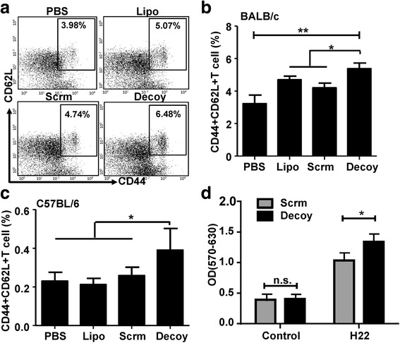
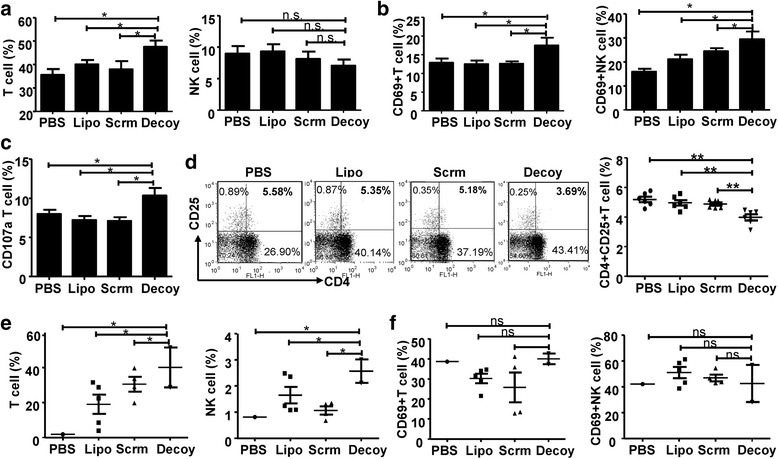
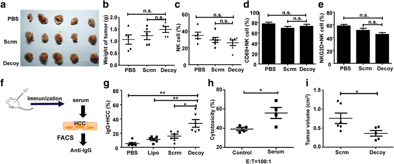
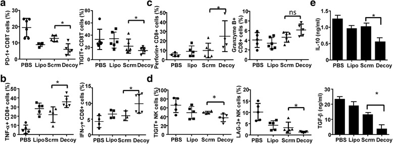
Results: STAT3-blocked whole HCC cell lysates inhibited tumor growth and tumorigenesis, and prolonged the survival of tumor-bearing mice. In addition, STAT3-blocked whole HCC cell lysates stimulated the activation of T cells and natural killer (NK) cells, and enhanced the infiltration of cytotoxic CD8+ T cells in the tumor tissues. In addition, the maturation of dendritic cells (DCs) was enhanced, which promoted the generation of immunological memory against HCC. Furthermore, secondary immune responses could be primed as soon as these immunized mice were challenged with HCC cells, accompanied by T cell and NK cell activation and infiltration. Additionally, immunization with this vaccine decreased the generation of Tregs and the production of TGF-β and IL-10. Importantly, STAT3-blocked whole HCC cell lysates prevented HCC-mediated exhaustion of T cells and NK cells, showing low expression of checkpoint molecules such as PD-1 and TIGIT on T cells and NK cells in the immunized mice.

Conclusions: The newly generated STAT3-blocked whole-cell HCC vaccine has potential for cancer cell vaccination.

Keywords: Hepatoma; Immunotherapy; STAT3; Tumor vaccine; Whole-cell vaccine.


Conflict of interest statement

Ethics approval and consent to participate

The animal research was approved by the Animal Research Ethics Committee of Shandong University, and complied with the Guidelines for Animal Experiments of Laboratory Animals.

Consent for publication

Not applicable.

Competing interests

The authors declare that they have no competing interests.

Publisher’s Note

Springer Nature remains neutral with regard to jurisdictional claims in published maps and institutional affiliations.

Figures

Fig. 1
Fig. 1
The HCC-vaccine could inhibit tumorigenesis and progression. BALB/c and C57BL/6 mice were immunized with H22- or Hepa1–6- cell derived tumor vaccine once a week for three weeks. At the first week after the last immunization, BALB/c and C57BL/6 mice were inoculated subcutaneously with 2 × 106 H22 or Hepa1–6 cells, respectively. The tumor volume growth curve of tumor-bearing BALB/c (a) and C57BL/6 (c) mice were monitored. b Tumor size and tumor weight of BALB/c were detected at week 4, mice in the PBS group had all died within 4 weeks. d The survival of tumor-bearing C57BL/6 mice immunized with HCC vaccine was monitored until all the mice died. PBS, mice immunized with PBS. Lipo, Scrm, and Decoy, mice immunized with Lipo, scrambled, or Decoy-ODN-treated HCC vaccine. Data are expressed as the mean ± SD, statistical significance was determined as *p < 0.05, **p < 0.01 and ***p < 0.001 (n = 5)
Fig. 2
Fig. 2
The HCC-vaccine activated the immune system in vivo. BALB/c mice were immunized with the H22 tumor vaccine once a week for three weeks. At the first week after the last immunization, PBMCs were isolated and analyzed by flow cytometry. a The proportion of CD3+DX5 or CD3DX5+ cells in PBMC of BALB/c were assayed. b & c The expression of CD69 on T cells and NK cells of BALB/c mice are shown (shaded histograms are isotype controls). d & e The proportion of CD11c+ DC cells and the expression of costimulatory molecules CD80 and CD86 on DCs were detected by FACS. PBS, mice immunized with PBS; Lipo, Scrm, and Decoy, mice immunized with Lipo, scrambled, or Decoy-ODN-treated HCC vaccine. Data are expressed as the mean ± SD, statistical significance was determined as *p < 0.05; **p < 0.01 and ***p < 0.001 (n ≥ 4)
Fig. 3
Fig. 3
The HCC vaccine promoted the generation of immune memory. PBMCs of BALB/c mice and C57BL/6 mice were isolated at the first week after the last immunization. The proportion of CD44+ CD62L+ cells in PBMCs from BALB/c mice (a & b) and C57BL/6 mice (c) was detected by FACS. d. The splenocytes of BALB/c mice were isolated at the first week after the last immunization, and an MTT assay was used to test the proliferation of the splenocytes treated or untreated (Control) with mitomycin-inactivated H22 cells for 5 days. PBS, mice immunized with PBS; Lipo, Scrm, and Decoy, mice immunized with Lipo, scrambled, or Decoy-ODN-treated HCC vaccine. Data are expressed as the mean ± SD, statistical significance was determined as *p < 0.05 and **p < 0.01 (n = 6)
Fig. 4
Fig. 4
The HCC vaccine induced a secondary immune response to HCC. BALB/c mice were inoculated subcutaneously with 2 × 106 tumor cells after three rounds of immunization with the HCC-vaccine, and then the PBMCs or tumor infiltrating lymphocytes of BALB/c mice were isolated when the tumor was visible. a The proportion of CD3+DX5 or CD3DX5+ cells in the PBMCs of BALB/c. b & c The expression of CD69 and CD107a on T cells or NK cells in the PBMCs of BALB/c were analyzed by flow cytometry. d The proportion of Tregs (CD3+CD4+CD25+) in the PBMCs was detected by flow cytometry. e & f The proportion and the expression of CD69 on T cells and NK cells in tumor tissues from BALB/c mice were analyzed by flow cytometry. Data are expressed as the mean ± SD, statistical significance was determined as *p < 0.05 and **p < 0.01 (n = 6)
Fig. 5
Fig. 5
The HCC vaccine-induced anti-tumor effect was dependent on cellular and humoral immunity. Nude mice were inoculated subcutaneously with 2 × 106 H22 cells after three rounds of immunization with the HCC vaccine. The tumor volume (a) and tumor weight (b) were detected at week 4. c-e PBMCs were isolated when the tumor was visible, and the proportion of NK cells (c), and the expression of CD69 (d) and NKG2D (e) on NK cells were detected by FACS. f-h Hepa1–6 cells were cultured with the serum from C57BL/6 mice immunized with the HCC vaccine for 4 h at 1% concentration. IgG+ cells were labeled by anti-mouse IgG antibody and analyzed by FACS (g); and, the cytotoxicity of spleen lymphocytes obtained from mice untreated with these Hepa1–6 cells was analyzed by 7-Aminoactinomycin D (7-AAD) staining methods (h). i BALB/c mice were immunized with H22 tumor vaccine once a week for three weeks. The PBMCs were isolated one week after the last immunization and transferred into healthy pre-irradiated BALB/c mice. After one week, these receptive mice were inoculated subcutaneously with 2 × 106 H22 and the tumor volumes were measured after four weeks. Data are expressed as the mean ± SD, statistical significance was determined as *p < 0.05 and ** p < 0.01 (n = 4–6)
Fig. 6
Fig. 6
The STAT3-blocked HCC-vaccine prevented tumor-induced CD8+ T and NK cell exhaustion. BALB/c mice were inoculated subcutaneously with 2 × 106 H22 cells after three rounds immunization. Four weeks later, the mice were killed when the control mice became moribund, and mononuclear cells were isolated from their spleens. a The levels of PD-1, CTLA-4, TIM-3, TIGIT, and LAG-3 were detected by FACS on T cells from the different groups. b Intracellular TNF-α and IFN-γ of CD4+ T cells were detected by FACS. c The percentage of Granzym B and perforin positive CD8+ T cells were assayed by FACS. d The levels of PD-1, CTLA-4, TIM-3, TIGIT and LAG-3 on NK cells was analyzed by FACS. e The levels of IL-10 and TGF-β in the serum of mice were assayed by enzyme-linked immunosorbent assay (ELISA). Data are expressed as the mean ± SD, statistical significance was determined as *p < 0.05 and **p < 0.01 (n = 6)

References

    1. Tagliamonte M, Petrizzo A, Tornesello ML, Ciliberto G, Buonaguro FM, Buonaguro L. Combinatorial immunotherapy strategies for hepatocellular carcinoma. Curr Opin Immunol. 2016;39:103–113. doi: 10.1016/j.coi.2016.01.005. - DOI - PubMed
    1. Mikhail S, Cosgrove D, Zeidan A. Hepatocellular carcinoma: systemic therapies and future perspectives. Expert Rev Anticancer Ther. 2014;14(10):1205–1218. doi: 10.1586/14737140.2014.949246. - DOI - PubMed
    1. Buonaguro L, Petrizzo A, Tagliamonte M, Tornesello ML, Buonaguro FM. Challenges in cancer vaccine development for hepatocellular carcinoma. J Hepatol. 2013;59(4):897–903. doi: 10.1016/j.jhep.2013.05.031. - DOI - PubMed
    1. Greten TF, Forner A, Korangy F, N'Kontchou G, Barget N, Ayuso C, Ormandy LA, Manns MP, Beaugrand M, Bruix J. A phase II open label trial evaluating safety and efficacy of a telomerase peptide vaccination in patients with advanced hepatocellular carcinoma. BMC Cancer. 2010;10:209. doi: 10.1186/1471-2407-10-209. - DOI - PMC - PubMed
    1. Saini N, Srinivasan R, Chawla Y, Sharma S, Chakraborti A, Rajwanshi A. Telomerase activity, telomere length and human telomerase reverse transcriptase expression in hepatocellular carcinoma is independent of hepatitis virus status. Liver Int. 2009;29(8):1162–1170. doi: 10.1111/j.1478-3231.2009.02082.x. - DOI - PubMed

Publication types

MeSH terms